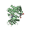
Entry Database : PDB / ID : 6sm8Title Human jak1 kinase domain in complex with inhibitor (Tyrosine-protein kinase JAK1) x 2 Keywords / Function / homology Function Domain/homology Component
/ / / / / / / / / / / / / / / / / / / / / / / / / / / / / / / / / / / / / / / / / / / / / / / / / / / / / / / / / / / / / / / / / / / / / / / / / / / / / / / / / / / / / / / / / / / / / / / / / / / / / / / / / / / / / / / / / / / / / / Biological species Homo sapiens (human)Method / / / Resolution : 1.85 Å Authors Read, J.A. / Steuber, H. Journal : J.Med.Chem. / Year : 2020Title : Discovery of (2R)-N-[3-[2-[(3-Methoxy-1-methyl-pyrazol-4-yl)amino]pyrimidin-4-yl]-1H-indol-7-yl]-2-(4-methylpiperazin-1-yl)propenamide (AZD4205) as a Potent and Selective Janus Kinase 1 Inhibitor.Authors: Su, Q. / Banks, E. / Bebernitz, G. / Bell, K. / Borenstein, C.F. / Chen, H. / Chuaqui, C.E. / Deng, N. / Ferguson, A.D. / Kawatkar, S. / Grimster, N.P. / Ruston, L. / Lyne, P.D. / Read, J.A. ... Authors : Su, Q. / Banks, E. / Bebernitz, G. / Bell, K. / Borenstein, C.F. / Chen, H. / Chuaqui, C.E. / Deng, N. / Ferguson, A.D. / Kawatkar, S. / Grimster, N.P. / Ruston, L. / Lyne, P.D. / Read, J.A. / Peng, X. / Pei, X. / Fawell, S. / Tang, Z. / Throner, S. / Vasbinder, M.M. / Wang, H. / Winter-Holt, J. / Woessner, R. / Wu, A. / Yang, W. / Zinda, M. / Kettle, J.G. History Deposition Aug 21, 2019 Deposition site / Processing site Revision 1.0 Apr 29, 2020 Provider / Type Revision 1.1 May 13, 2020 Group / Category / citation_authorItem / _citation_author.identifier_ORCID / _citation_author.nameRevision 1.2 May 27, 2020 Group / Category / citation_authorItem _citation.journal_volume / _citation.page_first ... _citation.journal_volume / _citation.page_first / _citation.page_last / _citation_author.identifier_ORCID Revision 1.3 Nov 6, 2024 Group Advisory / Data collection ... Advisory / Data collection / Database references / Structure summary Category chem_comp_atom / chem_comp_bond ... chem_comp_atom / chem_comp_bond / database_2 / pdbx_entry_details / pdbx_modification_feature / pdbx_unobs_or_zero_occ_atoms Item / _database_2.pdbx_database_accession / _pdbx_entry_details.has_protein_modification
Show all Show less
 Movie
Movie Controller


 Open data
 Basic information
 Components
 Keywords
 Function and homology information
 Homo sapiens (human)
 X-RAY DIFFRACTION /
  Authors
 Citation
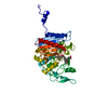
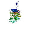
 Structure visualization
 Downloads & links
 Other downloads
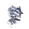


 PDBj
PDBj


 Assembly
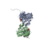


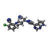
 Mass: 442.904 Da / Num. of mol.: 2 / Source method: obtained synthetically / Formula: C23H19ClN8 / Feature type: SUBJECT OF INVESTIGATION
 Mass: 62.068 Da / Num. of mol.: 2 / Source method: obtained synthetically / Formula: C2H6O2
 Mass: 18.015 Da / Num. of mol.: 254 / Source method: isolated from a natural source / Formula: H2O
 Sample preparation
 / Beamline: X06SA / Wavelength: 0.99999 Å
 Processing