 Movie
Movie Controller


+ Open data

- Basic information
| Entry | Database: PDB / ID: 6sjh | ||||||
|---|---|---|---|---|---|---|---|
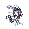
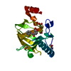
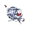
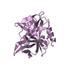
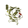
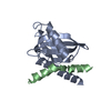
| Title | Structure of the PRY-SPRY domain of human Trim16L/Trim70 | ||||||
 Components Components | Tripartite motif-containing protein 16-like protein | ||||||
 Keywords Keywords | UNKNOWN FUNCTION / Unknown | ||||||
| Function / homology |  Function and homology information Function and homology information | ||||||
| Biological species |  Homo sapiens (human) Homo sapiens (human) | ||||||
| Method |  X-RAY DIFFRACTION / SYNCHROTRON / MOLECULAR REPLACEMENT / Resolution: 1.5 Å X-RAY DIFFRACTION / SYNCHROTRON / MOLECULAR REPLACEMENT / Resolution: 1.5 Å | ||||||
 Authors Authors | Mikkelsen, J.H. / Andersen, C.B.F. | ||||||
 Citation Citation | Journal: To Be Published Title: Structure of the PRY-SPRY domain of human Trim16L/Trim70 Authors: Mikkelsen, J.H. / Andersen, C.B.F. | ||||||
| History |
|
- Structure visualization
| Structure viewer | Molecule: MolmilJmol/JSmol |
|---|
- Downloads & links
-Download
| PDBx/mmCIF format | 6sjh.cif.gz | 257.4 KB | Display | PDBx/mmCIF format |
|---|---|---|---|---|
| PDB format | pdb6sjh.ent.gz | 205.9 KB | Display | PDB format |
| PDBx/mmJSON format | 6sjh.json.gz | Tree view | PDBx/mmJSON format | |
| Others |  Other downloads Other downloads |
-Validation report
| Arichive directory | https://data.pdbj.org/pub/pdb/validation_reports/sj/6sjhftp://data.pdbj.org/pub/pdb/validation_reports/sj/6sjh | HTTPS FTP |
|---|
-Related structure data
| Related structure data | 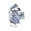 4b8eS S: Starting model for refinement |
|---|---|
| Similar structure data |


-Links
 PDBj
PDBj
- Assembly
| Deposited unit | 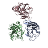
| ||||||||||||
|---|---|---|---|---|---|---|---|---|---|---|---|---|---|
| 1 | 
| ||||||||||||
| 2 | 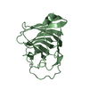
| ||||||||||||
| 3 | 
| ||||||||||||
| Unit cell |
|
-Components
| #1: Protein | Mass: 22315.129 Da / Num. of mol.: 3 Source method: isolated from a genetically manipulated source Source: (gene. exp.) Homo sapiens (human) / Gene: TRIM16L, TRIM70 / Production host:  #2: Water | ChemComp-HOH / |  Mass: 18.015 Da / Num. of mol.: 637 / Source method: isolated from a natural source / Formula: H2O Mass: 18.015 Da / Num. of mol.: 637 / Source method: isolated from a natural source / Formula: H2O |
|---|
-Experimental details
-Experiment
| Experiment | Method: X-RAY DIFFRACTION / Number of used crystals: 1 |
|---|
- Sample preparation
| Crystal | Density Matthews: 3.02 Å3/Da / Density % sol: 59.29 % |
|---|---|
| Crystal grow | Temperature: 277 K / Method: vapor diffusion, sitting drop / pH: 5 / Details: Na-acetate, PEG 400, PGA |
-Data collection
| Diffraction | Mean temperature: 100 K / Serial crystal experiment: N |
|---|---|
| Diffraction source | Source: SYNCHROTRON / Site: PETRA III, EMBL c/o DESY  / Beamline: P13 (MX1) / Wavelength: 0.9763 Å / Beamline: P13 (MX1) / Wavelength: 0.9763 Å |
| Detector | Type: DECTRIS PILATUS3 S 6M / Detector: PIXEL / Date: Jul 13, 2019 |
| Radiation | Protocol: SINGLE WAVELENGTH / Monochromatic (M) / Laue (L): M / Scattering type: x-ray |
| Radiation wavelength | Wavelength: 0.9763 Å / Relative weight: 1 |
| Reflection | Resolution: 1.5→30 Å / Num. obs: 221803 / % possible obs: 88.3 % / Redundancy: 2.48 % / CC1/2: 0.993 / Rmerge(I) obs: 0.075 / Net I/σ(I): 7.04 |
| Reflection shell | Resolution: 1.5→1.6 Å / Redundancy: 2.34 % / Num. unique obs: 95480 / CC1/2: 0.644 / Rrim(I) all: 0.555 / % possible all: 92.1 |
- Processing
| Software |
| ||||||||||||||||
|---|---|---|---|---|---|---|---|---|---|---|---|---|---|---|---|---|---|
| Refinement | Method to determine structure: MOLECULAR REPLACEMENT Starting model: 4B8E Resolution: 1.5→30 Å / Cross valid method: FREE R-VALUE
| ||||||||||||||||
| Refinement step | Cycle: LAST / Resolution: 1.5→30 Å
|
