 Movie
Movie Controller

[English] 日本語
 Yorodumi
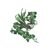
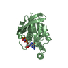
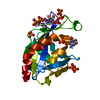
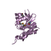
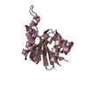
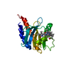
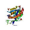
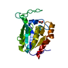
- PDB-1kmq: Crystal Structure of a Constitutively Activated RhoA Mutant (Q63L) -
+ Open data

- Basic information
| Entry | Database: PDB / ID: 1kmq | ||||||
|---|---|---|---|---|---|---|---|
| Title | Crystal Structure of a Constitutively Activated RhoA Mutant (Q63L) | ||||||
 Components Components | TRANSFORMING PROTEIN RHOA | ||||||
 Keywords Keywords | SIGNALING PROTEIN / GTPase / constitutive mutant / GTP-analog | ||||||
| Function / homology |  Function and homology information Function and homology informationalpha-beta T cell lineage commitment / aortic valve formation / beta selection / positive regulation of lipase activity / endothelial tube lumen extension / skeletal muscle satellite cell migration / bone trabecula morphogenesis / SLIT2:ROBO1 increases RHOA activity / RHO GTPases Activate Rhotekin and Rhophilins / Roundabout signaling pathway ...alpha-beta T cell lineage commitment / aortic valve formation / beta selection / positive regulation of lipase activity / endothelial tube lumen extension / skeletal muscle satellite cell migration / bone trabecula morphogenesis / SLIT2:ROBO1 increases RHOA activity / RHO GTPases Activate Rhotekin and Rhophilins / Roundabout signaling pathway / Axonal growth inhibition (RHOA activation) / Axonal growth stimulation / cleavage furrow formation / negative regulation of cell size / regulation of osteoblast proliferation / regulation of modification of postsynaptic actin cytoskeleton / mitotic cleavage furrow formation / apical junction assembly / negative regulation of cell migration involved in sprouting angiogenesis / establishment of epithelial cell apical/basal polarity / positive regulation of alpha-beta T cell differentiation / cell junction assembly / cellular response to chemokine / negative regulation of oxidative phosphorylation / regulation of modification of postsynaptic structure / RHO GTPases Activate ROCKs / RHO GTPases activate CIT / odontogenesis / PCP/CE pathway / Sema4D induced cell migration and growth-cone collapse / RHO GTPases activate KTN1 / apolipoprotein A-I-mediated signaling pathway / Sema4D mediated inhibition of cell attachment and migration / wound healing, spreading of cells / stress fiber assembly / positive regulation of leukocyte adhesion to vascular endothelial cell / Wnt signaling pathway, planar cell polarity pathway / PI3K/AKT activation / regulation of focal adhesion assembly / positive regulation of protein serine/threonine kinase activity / negative chemotaxis / ossification involved in bone maturation / EPHA-mediated growth cone collapse / apical junction complex / myosin binding / positive regulation of cytokinesis / RHOC GTPase cycle / cellular response to cytokine stimulus / ERBB2 Regulates Cell Motility / cleavage furrow / semaphorin-plexin signaling pathway / negative regulation of cell-substrate adhesion / mitotic spindle assembly / Rho protein signal transduction / ficolin-1-rich granule membrane / RHOA GTPase cycle / endothelial cell migration / positive regulation of stress fiber assembly / substrate adhesion-dependent cell spreading / positive regulation of T cell migration / positive regulation of neuron differentiation / GPVI-mediated activation cascade / RHO GTPases activate PKNs / negative regulation of reactive oxygen species biosynthetic process / PTK6 Regulates RHO GTPases, RAS GTPase and MAP kinases / cytoplasmic microtubule organization / EPHB-mediated forward signaling / regulation of cell migration / substantia nigra development / secretory granule membrane / regulation of actin cytoskeleton organization / cell periphery / small monomeric GTPase / TGF-beta receptor signaling in EMT (epithelial to mesenchymal transition) / regulation of microtubule cytoskeleton organization / RHO GTPases Activate Formins / positive regulation of non-canonical NF-kappaB signal transduction / cell junction / VEGFA-VEGFR2 Pathway / ruffle membrane / actin cytoskeleton organization / Ovarian tumor domain proteases / cell migration / G beta:gamma signalling through PI3Kgamma / lamellipodium / G alpha (12/13) signalling events / cellular response to lipopolysaccharide / midbody / G protein activity / cell cortex / cytoskeleton / dendritic spine / vesicle / cytoplasmic side of plasma membrane / positive regulation of canonical NF-kappaB signal transduction / postsynapse / endosome / focal adhesion / GTPase activity / Neutrophil degranulation Similarity search - Function | ||||||
| Biological species |  Homo sapiens (human) Homo sapiens (human) | ||||||
| Method |  X-RAY DIFFRACTION / SYNCHROTRON / MOLECULAR REPLACEMENT / Resolution: 1.55 Å X-RAY DIFFRACTION / SYNCHROTRON / MOLECULAR REPLACEMENT / Resolution: 1.55 Å | ||||||
 Authors Authors | Longenecker, K. / Read, P. / Lin, S.-K. / Somlyo, A.P. / Nakamoto, R.K. / Derewenda, Z.S. | ||||||
 Citation Citation | Journal: Acta Crystallogr.,Sect.D / Year: 2003 Title: Structure of a constitutively activated RhoA mutant (Q63L) at 1.55 A resolution. Authors: Longenecker, K. / Read, P. / Lin, S.K. / Somlyo, A.P. / Nakamoto, R.K. / Derewenda, Z.S. | ||||||
| History |
|
- Structure visualization
| Structure viewer | Molecule: MolmilJmol/JSmol |
|---|
- Downloads & links
-Download
| PDBx/mmCIF format | 1kmq.cif.gz | 95.1 KB | Display | PDBx/mmCIF format |
|---|---|---|---|---|
| PDB format | pdb1kmq.ent.gz | 70.7 KB | Display | PDB format |
| PDBx/mmJSON format | 1kmq.json.gz | Tree view | PDBx/mmJSON format | |
| Others |  Other downloads Other downloads |
-Validation report
| Arichive directory | https://data.pdbj.org/pub/pdb/validation_reports/km/1kmqftp://data.pdbj.org/pub/pdb/validation_reports/km/1kmq | HTTPS FTP |
|---|
-Related structure data
| Similar structure data |
|---|

-Links
 PDBj
PDBj








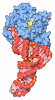

- Assembly
| Deposited unit | 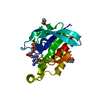
| ||||||||
|---|---|---|---|---|---|---|---|---|---|
| 1 |
| ||||||||
| Unit cell |
|
-Components
| #1: Protein | Mass: 20646.635 Da / Num. of mol.: 1 / Mutation: Q63L, F25N Source method: isolated from a genetically manipulated source Source: (gene. exp.) Homo sapiens (human) / Production host:   Escherichia coli (E. coli) / References: UniProt: P61586 Escherichia coli (E. coli) / References: UniProt: P61586 | ||||||
|---|---|---|---|---|---|---|---|
| #2: Chemical |   Mass: 24.305 Da / Num. of mol.: 2 / Source method: obtained synthetically / Formula: Mg Mass: 24.305 Da / Num. of mol.: 2 / Source method: obtained synthetically / Formula: Mg#3: Chemical | ChemComp-GNP / | 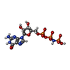 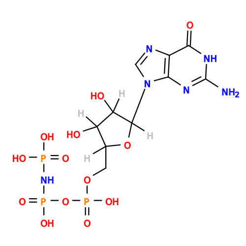 Mass: 522.196 Da / Num. of mol.: 1 / Source method: obtained synthetically / Formula: C10H17N6O13P3 Mass: 522.196 Da / Num. of mol.: 1 / Source method: obtained synthetically / Formula: C10H17N6O13P3Comment: GppNHp, GMPPNP, energy-carrying molecule analogue*YM #4: Chemical |  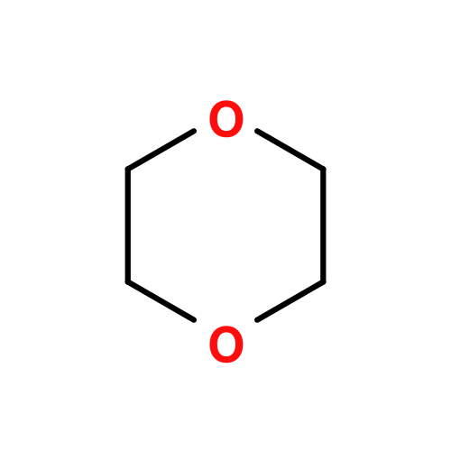 Mass: 88.105 Da / Num. of mol.: 2 / Source method: obtained synthetically / Formula: C4H8O2 Mass: 88.105 Da / Num. of mol.: 2 / Source method: obtained synthetically / Formula: C4H8O2#5: Water | ChemComp-HOH / |  Mass: 18.015 Da / Num. of mol.: 210 / Source method: isolated from a natural source / Formula: H2O Mass: 18.015 Da / Num. of mol.: 210 / Source method: isolated from a natural source / Formula: H2O |
-Experimental details
-Experiment
| Experiment | Method: X-RAY DIFFRACTION / Number of used crystals: 1 |
|---|
- Sample preparation
| Crystal | Density Matthews: 2.58 Å3/Da / Density % sol: 52.38 % | ||||||||||||||||||||||||||||||
|---|---|---|---|---|---|---|---|---|---|---|---|---|---|---|---|---|---|---|---|---|---|---|---|---|---|---|---|---|---|---|---|
| Crystal grow | Temperature: 282 K / Method: vapor diffusion / pH: 6.9 Details: PEG 8000, HEPES, dioxane, magnesium chloride, 2-mercaptoethanol, pH 6.9, VAPOR DIFFUSION, temperature 282K | ||||||||||||||||||||||||||||||
| Crystal grow | *PLUS Temperature: 277 K | ||||||||||||||||||||||||||||||
| Components of the solutions | *PLUS
|
-Data collection
| Diffraction | Mean temperature: 100 K |
|---|---|
| Diffraction source | Source: SYNCHROTRON / Site: NSLS  / Beamline: X9B / Wavelength: 0.9671 Å / Beamline: X9B / Wavelength: 0.9671 Å |
| Detector | Type: ADSC QUANTUM 4 / Detector: CCD / Date: Jun 19, 2001 |
| Radiation | Protocol: SINGLE WAVELENGTH / Monochromatic (M) / Laue (L): M / Scattering type: x-ray |
| Radiation wavelength | Wavelength: 0.9671 Å / Relative weight: 1 |
| Reflection | Resolution: 1.55→30 Å / Num. obs: 30966 / % possible obs: 97.3 % / Observed criterion σ(F): 0 / Redundancy: 5 % / Rsym value: 0.036 / Net I/σ(I): 18.9 |
| Reflection shell | Resolution: 1.55→1.61 Å / Mean I/σ(I) obs: 5.4 / Rsym value: 0.28 / % possible all: 84.6 |
| Reflection | *PLUS Lowest resolution: 30 Å / % possible obs: 97 % / Num. measured all: 154149 / Rmerge(I) obs: 0.036 |
| Reflection shell | *PLUS % possible obs: 85 % / Rmerge(I) obs: 0.28 |
- Processing
| Software |
| ||||||||||||||||||||||||||||||||||||||||||||||||||||||||||||||||||||||||||||||||||||||||||||||||||||||||||||||||||||||||||||||||||
|---|---|---|---|---|---|---|---|---|---|---|---|---|---|---|---|---|---|---|---|---|---|---|---|---|---|---|---|---|---|---|---|---|---|---|---|---|---|---|---|---|---|---|---|---|---|---|---|---|---|---|---|---|---|---|---|---|---|---|---|---|---|---|---|---|---|---|---|---|---|---|---|---|---|---|---|---|---|---|---|---|---|---|---|---|---|---|---|---|---|---|---|---|---|---|---|---|---|---|---|---|---|---|---|---|---|---|---|---|---|---|---|---|---|---|---|---|---|---|---|---|---|---|---|---|---|---|---|---|---|---|---|
| Refinement | Method to determine structure: MOLECULAR REPLACEMENT / Resolution: 1.55→48 Å / Cor.coef. Fo:Fc: 0.964 / Cor.coef. Fo:Fc free: 0.957 / SU B: 3.121 / SU ML: 0.059 / Cross valid method: THROUGHOUT / σ(F): 0 / ESU R: 0.086 / ESU R Free: 0.072 / Stereochemistry target values: Engh & Huber
| ||||||||||||||||||||||||||||||||||||||||||||||||||||||||||||||||||||||||||||||||||||||||||||||||||||||||||||||||||||||||||||||||||
| Solvent computation | Ion probe radii: 0.8 Å / Shrinkage radii: 0.8 Å / VDW probe radii: 1.4 Å | ||||||||||||||||||||||||||||||||||||||||||||||||||||||||||||||||||||||||||||||||||||||||||||||||||||||||||||||||||||||||||||||||||
| Displacement parameters | Biso mean: 20.907 Å2
| ||||||||||||||||||||||||||||||||||||||||||||||||||||||||||||||||||||||||||||||||||||||||||||||||||||||||||||||||||||||||||||||||||
| Refinement step | Cycle: LAST / Resolution: 1.55→48 Å
| ||||||||||||||||||||||||||||||||||||||||||||||||||||||||||||||||||||||||||||||||||||||||||||||||||||||||||||||||||||||||||||||||||
| Refine LS restraints |
| ||||||||||||||||||||||||||||||||||||||||||||||||||||||||||||||||||||||||||||||||||||||||||||||||||||||||||||||||||||||||||||||||||
| LS refinement shell | Resolution: 1.55→1.59 Å / Total num. of bins used: 20 /
| ||||||||||||||||||||||||||||||||||||||||||||||||||||||||||||||||||||||||||||||||||||||||||||||||||||||||||||||||||||||||||||||||||
| Software | *PLUS Version: 5 / Classification: refinement | ||||||||||||||||||||||||||||||||||||||||||||||||||||||||||||||||||||||||||||||||||||||||||||||||||||||||||||||||||||||||||||||||||
| Refinement | *PLUS % reflection Rfree: 5 % / Rfactor Rfree: 0.19 / Rfactor Rwork: 0.161 | ||||||||||||||||||||||||||||||||||||||||||||||||||||||||||||||||||||||||||||||||||||||||||||||||||||||||||||||||||||||||||||||||||
| Solvent computation | *PLUS | ||||||||||||||||||||||||||||||||||||||||||||||||||||||||||||||||||||||||||||||||||||||||||||||||||||||||||||||||||||||||||||||||||
| Displacement parameters | *PLUS | ||||||||||||||||||||||||||||||||||||||||||||||||||||||||||||||||||||||||||||||||||||||||||||||||||||||||||||||||||||||||||||||||||
| Refine LS restraints | *PLUS
|
