 ムービー
ムービー コントローラー


+ データを開く

- 基本情報
基本情報
| 登録情報 | データベース: PDB / ID: 6sj5 | ||||||
|---|---|---|---|---|---|---|---|
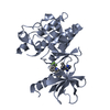
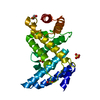
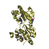
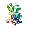
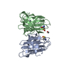
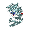
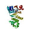
| タイトル | Crystal structure of the uL14-RsfS complex from Staphylococcus aureus | ||||||
 要素 要素 |
| ||||||
 キーワード キーワード | RIBOSOME / Stress / S.aureus / RsfS / Hibernation | ||||||
| 機能・相同性 |  機能・相同性情報 機能・相同性情報negative regulation of ribosome biogenesis / cytosolic ribosome assembly / large ribosomal subunit rRNA binding / cytosolic large ribosomal subunit / negative regulation of translation / structural constituent of ribosome / translation / cytoplasm 類似検索 - 分子機能 | ||||||
| 生物種 |   Staphylococcus aureus subsp. aureus (黄色ブドウ球菌)Staphylococcus aureus subsp. aureus NCTC 8325 (黄色ブドウ球菌) Staphylococcus aureus subsp. aureus (黄色ブドウ球菌)Staphylococcus aureus subsp. aureus NCTC 8325 (黄色ブドウ球菌) | ||||||
| 手法 |  X線回折 / シンクロトロン / 分子置換 / 解像度: 2.26686551714 Å X線回折 / シンクロトロン / 分子置換 / 解像度: 2.26686551714 Å | ||||||
 データ登録者 データ登録者 | Fatkhullin, B. / Gabdulkhakov, A. / Yusupova, G. / Yusupov, M. | ||||||
 引用 引用 | ジャーナル: Nat Commun / 年: 2020 タイトル: Mechanism of ribosome shutdown by RsfS in Staphylococcus aureus revealed by integrative structural biology approach. 著者: Iskander Khusainov / Bulat Fatkhullin / Simone Pellegrino / Aydar Bikmullin / Wen-Ti Liu / Azat Gabdulkhakov / Amr Al Shebel / Alexander Golubev / Denis Zeyer / Natalie Trachtmann / Georg A ...著者: Iskander Khusainov / Bulat Fatkhullin / Simone Pellegrino / Aydar Bikmullin / Wen-Ti Liu / Azat Gabdulkhakov / Amr Al Shebel / Alexander Golubev / Denis Zeyer / Natalie Trachtmann / Georg A Sprenger / Shamil Validov / Konstantin Usachev / Gulnara Yusupova / Marat Yusupov /     要旨: For the sake of energy preservation, bacteria, upon transition to stationary phase, tone down their protein synthesis. This process is favored by the reversible binding of small stress-induced ...For the sake of energy preservation, bacteria, upon transition to stationary phase, tone down their protein synthesis. This process is favored by the reversible binding of small stress-induced proteins to the ribosome to prevent unnecessary translation. One example is the conserved bacterial ribosome silencing factor (RsfS) that binds to uL14 protein onto the large ribosomal subunit and prevents its association with the small subunit. Here we describe the binding mode of Staphylococcus aureus RsfS to the large ribosomal subunit and present a 3.2 Å resolution cryo-EM reconstruction of the 50S-RsfS complex together with the crystal structure of uL14-RsfS complex solved at 2.3 Å resolution. The understanding of the detailed landscape of RsfS-uL14 interactions within the ribosome shed light on the mechanism of ribosome shutdown in the human pathogen S. aureus and might deliver a novel target for pharmacological drug development and treatment of bacterial infections. | ||||||
| 履歴 |
|
- 構造の表示
構造の表示
| 構造ビューア | 分子: MolmilJmol/JSmol |
|---|
- ダウンロードとリンク
-ダウンロード
| PDBx/mmCIF形式 | 6sj5.cif.gz | 220.8 KB | 表示 | PDBx/mmCIF形式 |
|---|---|---|---|---|
| PDB形式 | pdb6sj5.ent.gz | 159 KB | 表示 | PDB形式 |
| PDBx/mmJSON形式 | 6sj5.json.gz | ツリー表示 | PDBx/mmJSON形式 | |
| その他 |  その他のダウンロード その他のダウンロード |
-検証レポート
| 文書・要旨 | 6sj5_validation.pdf.gz | 481.4 KB | 表示 | wwPDB検証レポート |
|---|---|---|---|---|
| 文書・詳細版 | 6sj5_full_validation.pdf.gz | 491.5 KB | 表示 | |
| XML形式データ | 6sj5_validation.xml.gz | 19.3 KB | 表示 | |
| CIF形式データ | 6sj5_validation.cif.gz | 25.4 KB | 表示 | |
| アーカイブディレクトリ | https://data.pdbj.org/pub/pdb/validation_reports/sj/6sj5ftp://data.pdbj.org/pub/pdb/validation_reports/sj/6sj5 | HTTPS FTP |
-関連構造データ


-リンク
 PDBj
PDBj


- 集合体
集合体
| 登録構造単位 | 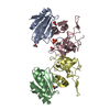
| ||||||||||||
|---|---|---|---|---|---|---|---|---|---|---|---|---|---|
| 1 | 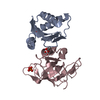
| ||||||||||||
| 2 | 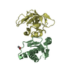
| ||||||||||||
| 単位格子 |
|
-要素
| #1: タンパク質 | 分子量: 13467.043 Da / 分子数: 2 / 由来タイプ: 組換発現 由来: (組換発現) Staphylococcus aureus subsp. aureus (黄色ブドウ球菌)遺伝子: rsfS, E5491_08675, FAF14_04505, FAF30_12305, FAF31_00945, FAF32_10770, QU38_08975 発現宿主: #2: タンパク質 | 分子量: 15328.716 Da / 分子数: 2 / 由来タイプ: 組換発現 由来: (組換発現) Staphylococcus aureus subsp. aureus NCTC 8325 (黄色ブドウ球菌)遺伝子: rplN, SAOUHSC_02502 / 発現宿主: #3: 化合物 | ChemComp-ACY /   分子量: 60.052 Da / 分子数: 4 / 由来タイプ: 合成 / 式: C2H4O2 分子量: 60.052 Da / 分子数: 4 / 由来タイプ: 合成 / 式: C2H4O2#4: 化合物 | ChemComp-PO4 / |   分子量: 94.971 Da / 分子数: 1 / 由来タイプ: 合成 / 式: PO4 分子量: 94.971 Da / 分子数: 1 / 由来タイプ: 合成 / 式: PO4#5: 水 | ChemComp-HOH / |  分子量: 18.015 Da / 分子数: 16 / 由来タイプ: 天然 / 式: H2O 分子量: 18.015 Da / 分子数: 16 / 由来タイプ: 天然 / 式: H2O研究の焦点であるリガンドがあるか | N | |
|---|
-実験情報
-実験
| 実験 | 手法: X線回折 / 使用した結晶の数: 1 |
|---|
- 試料調製
試料調製
| 結晶 | マシュー密度: 2.52 Å3/Da / 溶媒含有率: 51.23 % |
|---|---|
| 結晶化 | 温度: 295 K / 手法: 蒸気拡散法, ハンギングドロップ法 / pH: 6 詳細: 0.2 M Lithium sulfate, 0.1 M MES pH 6.0, 20% PEG 4000 |
-データ収集
| 回折 | 平均測定温度: 100 K / Serial crystal experiment: N |
|---|---|
| 放射光源 | 由来: シンクロトロン / サイト: ESRF / ビームライン: ID30B / 波長: 0.9762 Å |
| 検出器 | タイプ: DECTRIS PILATUS3 6M / 検出器: PIXEL / 日付: 2018年10月27日 |
| 放射 | プロトコル: SINGLE WAVELENGTH / 単色(M)・ラウエ(L): M / 散乱光タイプ: x-ray |
| 放射波長 | 波長: 0.9762 Å / 相対比: 1 |
| 反射 | 解像度: 2.267→42.29 Å / Num. obs: 25254 / % possible obs: 98.95 % / 冗長度: 4.48 % / Biso Wilson estimate: 69.5777354322 Å2 / CC1/2: 0.999 / Net I/σ(I): 14.53 |
| 反射 シェル | 解像度: 2.267→2.348 Å / 冗長度: 4.39 % / Mean I/σ(I) obs: 0.9 / Num. unique obs: 2494 / CC1/2: 0.273 / % possible all: 99.17 |
- 解析
解析
| ソフトウェア |
| ||||||||||||||||||||||||||||||||||||||||||||||||||||||||||||||||||||||
|---|---|---|---|---|---|---|---|---|---|---|---|---|---|---|---|---|---|---|---|---|---|---|---|---|---|---|---|---|---|---|---|---|---|---|---|---|---|---|---|---|---|---|---|---|---|---|---|---|---|---|---|---|---|---|---|---|---|---|---|---|---|---|---|---|---|---|---|---|---|---|---|
| 精密化 | 構造決定の手法: 分子置換 開始モデル: 2O5A, 5ND8 解像度: 2.26686551714→42.2029407831 Å / SU ML: 0.390702518887 / 交差検証法: FREE R-VALUE / σ(F): 1.35468090639 / 位相誤差: 32.7644601701 立体化学のターゲット値: GeoStd + Monomer Library + CDL v1.2
| ||||||||||||||||||||||||||||||||||||||||||||||||||||||||||||||||||||||
| 溶媒の処理 | 減衰半径: 0.9 Å / VDWプローブ半径: 1.11 Å / 溶媒モデル: FLAT BULK SOLVENT MODEL | ||||||||||||||||||||||||||||||||||||||||||||||||||||||||||||||||||||||
| 原子変位パラメータ | Biso mean: 88.2172954769 Å2 | ||||||||||||||||||||||||||||||||||||||||||||||||||||||||||||||||||||||
| 精密化ステップ | サイクル: LAST / 解像度: 2.26686551714→42.2029407831 Å
| ||||||||||||||||||||||||||||||||||||||||||||||||||||||||||||||||||||||
| 拘束条件 |
| ||||||||||||||||||||||||||||||||||||||||||||||||||||||||||||||||||||||
| LS精密化 シェル |
| ||||||||||||||||||||||||||||||||||||||||||||||||||||||||||||||||||||||
| 精密化 TLS | 手法: refined / Origin x: 5.82612513189 Å / Origin y: 3.81488691363 Å / Origin z: 13.3497025537 Å
| ||||||||||||||||||||||||||||||||||||||||||||||||||||||||||||||||||||||
| 精密化 TLSグループ | Selection details: all |
