 Movie
Movie Controller

[English] 日本語
 Yorodumi
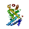
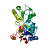
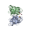
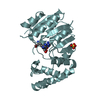
- PDB-6sj5: Crystal structure of the uL14-RsfS complex from Staphylococcus aureus -
+ Open data

- Basic information
| Entry | Database: PDB / ID: 6sj5 | ||||||
|---|---|---|---|---|---|---|---|
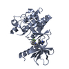
| Title | Crystal structure of the uL14-RsfS complex from Staphylococcus aureus | ||||||
 Components Components |
| ||||||
 Keywords Keywords | RIBOSOME / Stress / S.aureus / RsfS / Hibernation | ||||||
| Function / homology |  Function and homology information Function and homology informationnegative regulation of ribosome biogenesis / cytosolic ribosome assembly / large ribosomal subunit rRNA binding / cytosolic large ribosomal subunit / negative regulation of translation / structural constituent of ribosome / translation / cytoplasm Similarity search - Function | ||||||
| Biological species |   Staphylococcus aureus subsp. aureus (bacteria)Staphylococcus aureus subsp. aureus NCTC 8325 (bacteria) Staphylococcus aureus subsp. aureus (bacteria)Staphylococcus aureus subsp. aureus NCTC 8325 (bacteria) | ||||||
| Method |  X-RAY DIFFRACTION / SYNCHROTRON / MOLECULAR REPLACEMENT / Resolution: 2.26686551714 Å X-RAY DIFFRACTION / SYNCHROTRON / MOLECULAR REPLACEMENT / Resolution: 2.26686551714 Å | ||||||
 Authors Authors | Fatkhullin, B. / Gabdulkhakov, A. / Yusupova, G. / Yusupov, M. | ||||||
 Citation Citation | Journal: Nat Commun / Year: 2020 Title: Mechanism of ribosome shutdown by RsfS in Staphylococcus aureus revealed by integrative structural biology approach. Authors: Iskander Khusainov / Bulat Fatkhullin / Simone Pellegrino / Aydar Bikmullin / Wen-Ti Liu / Azat Gabdulkhakov / Amr Al Shebel / Alexander Golubev / Denis Zeyer / Natalie Trachtmann / Georg A ...Authors: Iskander Khusainov / Bulat Fatkhullin / Simone Pellegrino / Aydar Bikmullin / Wen-Ti Liu / Azat Gabdulkhakov / Amr Al Shebel / Alexander Golubev / Denis Zeyer / Natalie Trachtmann / Georg A Sprenger / Shamil Validov / Konstantin Usachev / Gulnara Yusupova / Marat Yusupov /     Abstract: For the sake of energy preservation, bacteria, upon transition to stationary phase, tone down their protein synthesis. This process is favored by the reversible binding of small stress-induced ...For the sake of energy preservation, bacteria, upon transition to stationary phase, tone down their protein synthesis. This process is favored by the reversible binding of small stress-induced proteins to the ribosome to prevent unnecessary translation. One example is the conserved bacterial ribosome silencing factor (RsfS) that binds to uL14 protein onto the large ribosomal subunit and prevents its association with the small subunit. Here we describe the binding mode of Staphylococcus aureus RsfS to the large ribosomal subunit and present a 3.2 Å resolution cryo-EM reconstruction of the 50S-RsfS complex together with the crystal structure of uL14-RsfS complex solved at 2.3 Å resolution. The understanding of the detailed landscape of RsfS-uL14 interactions within the ribosome shed light on the mechanism of ribosome shutdown in the human pathogen S. aureus and might deliver a novel target for pharmacological drug development and treatment of bacterial infections. | ||||||
| History |
|
- Structure visualization
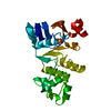
| Structure viewer | Molecule: MolmilJmol/JSmol |
|---|
- Downloads & links
-Download
| PDBx/mmCIF format | 6sj5.cif.gz | 220.8 KB | Display | PDBx/mmCIF format |
|---|---|---|---|---|
| PDB format | pdb6sj5.ent.gz | 159 KB | Display | PDB format |
| PDBx/mmJSON format | 6sj5.json.gz | Tree view | PDBx/mmJSON format | |
| Others |  Other downloads Other downloads |
-Validation report
| Arichive directory | https://data.pdbj.org/pub/pdb/validation_reports/sj/6sj5ftp://data.pdbj.org/pub/pdb/validation_reports/sj/6sj5 | HTTPS FTP |
|---|
-Related structure data
| Related structure data | 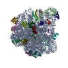 6sj6C  2o5aS 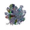 5nd8S S: Starting model for refinement C: citing same article ( |
|---|---|
| Similar structure data |


-Links
 PDBj
PDBj


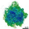
- Assembly
| Deposited unit | 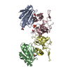
| ||||||||||||
|---|---|---|---|---|---|---|---|---|---|---|---|---|---|
| 1 | 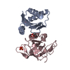
| ||||||||||||
| 2 | 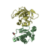
| ||||||||||||
| Unit cell |
|
-Components
| #1: Protein | Mass: 13467.043 Da / Num. of mol.: 2 Source method: isolated from a genetically manipulated source Source: (gene. exp.) Staphylococcus aureus subsp. aureus (bacteria)Gene: rsfS, E5491_08675, FAF14_04505, FAF30_12305, FAF31_00945, FAF32_10770, QU38_08975 Production host: #2: Protein | Mass: 15328.716 Da / Num. of mol.: 2 Source method: isolated from a genetically manipulated source Source: (gene. exp.) Staphylococcus aureus subsp. aureus NCTC 8325 (bacteria)Gene: rplN, SAOUHSC_02502 / Production host: #3: Chemical | ChemComp-ACY /   Mass: 60.052 Da / Num. of mol.: 4 / Source method: obtained synthetically / Formula: C2H4O2 Mass: 60.052 Da / Num. of mol.: 4 / Source method: obtained synthetically / Formula: C2H4O2#4: Chemical | ChemComp-PO4 / |   Mass: 94.971 Da / Num. of mol.: 1 / Source method: obtained synthetically / Formula: PO4 Mass: 94.971 Da / Num. of mol.: 1 / Source method: obtained synthetically / Formula: PO4#5: Water | ChemComp-HOH / |  Mass: 18.015 Da / Num. of mol.: 16 / Source method: isolated from a natural source / Formula: H2O Mass: 18.015 Da / Num. of mol.: 16 / Source method: isolated from a natural source / Formula: H2OHas ligand of interest | N | |
|---|
-Experimental details
-Experiment
| Experiment | Method: X-RAY DIFFRACTION / Number of used crystals: 1 |
|---|
- Sample preparation
| Crystal | Density Matthews: 2.52 Å3/Da / Density % sol: 51.23 % |
|---|---|
| Crystal grow | Temperature: 295 K / Method: vapor diffusion, hanging drop / pH: 6 Details: 0.2 M Lithium sulfate, 0.1 M MES pH 6.0, 20% PEG 4000 |
-Data collection
| Diffraction | Mean temperature: 100 K / Serial crystal experiment: N |
|---|---|
| Diffraction source | Source: SYNCHROTRON / Site: ESRF / Beamline: ID30B / Wavelength: 0.9762 Å |
| Detector | Type: DECTRIS PILATUS3 6M / Detector: PIXEL / Date: Oct 27, 2018 |
| Radiation | Protocol: SINGLE WAVELENGTH / Monochromatic (M) / Laue (L): M / Scattering type: x-ray |
| Radiation wavelength | Wavelength: 0.9762 Å / Relative weight: 1 |
| Reflection | Resolution: 2.267→42.29 Å / Num. obs: 25254 / % possible obs: 98.95 % / Redundancy: 4.48 % / Biso Wilson estimate: 69.5777354322 Å2 / CC1/2: 0.999 / Net I/σ(I): 14.53 |
| Reflection shell | Resolution: 2.267→2.348 Å / Redundancy: 4.39 % / Mean I/σ(I) obs: 0.9 / Num. unique obs: 2494 / CC1/2: 0.273 / % possible all: 99.17 |
- Processing
| Software |
| ||||||||||||||||||||||||||||||||||||||||||||||||||||||||||||||||||||||
|---|---|---|---|---|---|---|---|---|---|---|---|---|---|---|---|---|---|---|---|---|---|---|---|---|---|---|---|---|---|---|---|---|---|---|---|---|---|---|---|---|---|---|---|---|---|---|---|---|---|---|---|---|---|---|---|---|---|---|---|---|---|---|---|---|---|---|---|---|---|---|---|
| Refinement | Method to determine structure: MOLECULAR REPLACEMENT Starting model: 2O5A, 5ND8 Resolution: 2.26686551714→42.2029407831 Å / SU ML: 0.390702518887 / Cross valid method: FREE R-VALUE / σ(F): 1.35468090639 / Phase error: 32.7644601701 Stereochemistry target values: GeoStd + Monomer Library + CDL v1.2
| ||||||||||||||||||||||||||||||||||||||||||||||||||||||||||||||||||||||
| Solvent computation | Shrinkage radii: 0.9 Å / VDW probe radii: 1.11 Å / Solvent model: FLAT BULK SOLVENT MODEL | ||||||||||||||||||||||||||||||||||||||||||||||||||||||||||||||||||||||
| Displacement parameters | Biso mean: 88.2172954769 Å2 | ||||||||||||||||||||||||||||||||||||||||||||||||||||||||||||||||||||||
| Refinement step | Cycle: LAST / Resolution: 2.26686551714→42.2029407831 Å
| ||||||||||||||||||||||||||||||||||||||||||||||||||||||||||||||||||||||
| Refine LS restraints |
| ||||||||||||||||||||||||||||||||||||||||||||||||||||||||||||||||||||||
| LS refinement shell |
| ||||||||||||||||||||||||||||||||||||||||||||||||||||||||||||||||||||||
| Refinement TLS params. | Method: refined / Origin x: 5.82612513189 Å / Origin y: 3.81488691363 Å / Origin z: 13.3497025537 Å
| ||||||||||||||||||||||||||||||||||||||||||||||||||||||||||||||||||||||
| Refinement TLS group | Selection details: all |
