 Movie
Movie Controller

[English] 日本語
 Yorodumi
- PDB-6sh7: Crystal structure of the human DEAH-helicase DHX15 in complex wit... -
+ Open data

- Basic information
| Entry | Database: PDB / ID: 6sh7 | |||||||||
|---|---|---|---|---|---|---|---|---|---|---|
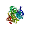
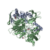
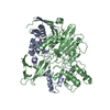
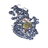
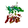
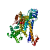
| Title | Crystal structure of the human DEAH-helicase DHX15 in complex with the NKRF G-patch | |||||||||
 Components Components |
| |||||||||
 Keywords Keywords | HYDROLASE / G-patch / DEAH helicase / DExH helicase | |||||||||
| Function / homology |  Function and homology information Function and homology informationU12-type spliceosomal complex / response to alkaloid / ATP-dependent activity, acting on RNA / ATPase activator activity / mRNA Splicing - Major Pathway / RNA splicing / antiviral innate immune response / helicase activity / spliceosomal complex / mRNA splicing, via spliceosome ...U12-type spliceosomal complex / response to alkaloid / ATP-dependent activity, acting on RNA / ATPase activator activity / mRNA Splicing - Major Pathway / RNA splicing / antiviral innate immune response / helicase activity / spliceosomal complex / mRNA splicing, via spliceosome / response to toxic substance / mRNA processing / double-stranded RNA binding / DNA-binding transcription activator activity, RNA polymerase II-specific / defense response to virus / positive regulation of canonical NF-kappaB signal transduction / RNA helicase activity / defense response to bacterium / nuclear speck / RNA helicase / RNA polymerase II cis-regulatory region sequence-specific DNA binding / negative regulation of DNA-templated transcription / nucleolus / DNA-templated transcription / positive regulation of transcription by RNA polymerase II / ATP hydrolysis activity / RNA binding / nucleoplasm / ATP binding / nucleus Similarity search - Function | |||||||||
| Biological species |  Homo sapiens (human) Homo sapiens (human) | |||||||||
| Method |  X-RAY DIFFRACTION / SYNCHROTRON / MOLECULAR REPLACEMENT / Resolution: 2.21 Å X-RAY DIFFRACTION / SYNCHROTRON / MOLECULAR REPLACEMENT / Resolution: 2.21 Å | |||||||||
 Authors Authors | Jonas, S. / Studer, M.K. / Ivanovic, L. | |||||||||
| Funding support |  Switzerland, 2items Switzerland, 2items
| |||||||||
 Citation Citation | Journal: Proc.Natl.Acad.Sci.USA / Year: 2020 Title: Structural basis for DEAH-helicase activation by G-patch proteins. Authors: Studer, M.K. / Ivanovic, L. / Weber, M.E. / Marti, S. / Jonas, S. #1: Journal: Acta Crystallogr. D Biol. Crystallogr. / Year: 2012 Title: Towards automated crystallographic structure refinement with phenix.refine. Authors: Afonine, P.V. / Grosse-Kunstleve, R.W. / Echols, N. / Headd, J.J. / Moriarty, N.W. / Mustyakimov, M. / Terwilliger, T.C. / Urzhumtsev, A. / Zwart, P.H. / Adams, P.D. | |||||||||
| History |
|
- Structure visualization
| Structure viewer | Molecule: MolmilJmol/JSmol |
|---|
- Downloads & links
-Download
| PDBx/mmCIF format | 6sh7.cif.gz | 358 KB | Display | PDBx/mmCIF format |
|---|---|---|---|---|
| PDB format | pdb6sh7.ent.gz | 244.5 KB | Display | PDB format |
| PDBx/mmJSON format | 6sh7.json.gz | Tree view | PDBx/mmJSON format | |
| Others |  Other downloads Other downloads |
-Validation report
| Arichive directory | https://data.pdbj.org/pub/pdb/validation_reports/sh/6sh7ftp://data.pdbj.org/pub/pdb/validation_reports/sh/6sh7 | HTTPS FTP |
|---|
-Related structure data
| Related structure data |  6sh6C  5xdrS C: citing same article ( S: Starting model for refinement |
|---|---|
| Similar structure data |
-Links
 PDBj
PDBj


- Assembly
| Deposited unit | 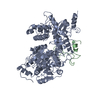
| ||||||||||||
|---|---|---|---|---|---|---|---|---|---|---|---|---|---|
| 1 |
| ||||||||||||
| Unit cell |
| ||||||||||||
| Components on special symmetry positions |
|
-Components
| #1: Protein | Mass: 79063.750 Da / Num. of mol.: 1 Source method: isolated from a genetically manipulated source Source: (gene. exp.) Homo sapiens (human) / Gene: DHX15, DBP1, DDX15 / Production host:  |
|---|---|
| #2: Protein | Mass: 7425.467 Da / Num. of mol.: 1 Source method: isolated from a genetically manipulated source Source: (gene. exp.) Homo sapiens (human) / Gene: NKRF, ITBA4, NRF / Production host: |
| #3: Water | ChemComp-HOH /  Mass: 18.015 Da / Num. of mol.: 34 / Source method: isolated from a natural source / Formula: H2O Mass: 18.015 Da / Num. of mol.: 34 / Source method: isolated from a natural source / Formula: H2O |
-Experimental details
-Experiment
| Experiment | Method: X-RAY DIFFRACTION / Number of used crystals: 1 |
|---|
- Sample preparation
| Crystal | Density Matthews: 2.43 Å3/Da / Density % sol: 49.37 % |
|---|---|
| Crystal grow | Temperature: 291.15 K / Method: vapor diffusion, sitting drop / pH: 8.5 Details: 0.1 M carboxylic acids, 0.1 M BICINE/Tris pH 8.5, 12.5% w/v PEG 1000, 12.5% w/v PEG 3350, 12.5% v/v MPD |
-Data collection
| Diffraction | Mean temperature: 100 K / Serial crystal experiment: N |
|---|---|
| Diffraction source | Source: SYNCHROTRON / Site: SLS / Beamline: X06SA / Wavelength: 1.000041 Å |
| Detector | Type: DECTRIS EIGER X 16M / Detector: PIXEL / Date: May 4, 2019 |
| Radiation | Protocol: SINGLE WAVELENGTH / Monochromatic (M) / Laue (L): M / Scattering type: x-ray |
| Radiation wavelength | Wavelength: 1.000041 Å / Relative weight: 1 |
| Reflection | Resolution: 2.21→46.23 Å / Num. obs: 39975 / % possible obs: 99.8 % / Redundancy: 13.1 % / Biso Wilson estimate: 61.95 Å2 / CC1/2: 0.998 / Rpim(I) all: 0.052 / Rrim(I) all: 0.187 / Net I/σ(I): 8.2 |
| Reflection shell | Resolution: 2.21→2.29 Å / Redundancy: 13.5 % / Num. unique obs: 3912 / CC1/2: 0.38 / Rpim(I) all: 0.763 / % possible all: 98.7 |
- Processing
| Software |
| |||||||||||||||||||||||||||||||||||||||||||||||||||||||||||||||||||||||||||||||||||||||||||||||||||||||||||||||||||||||||||||||||||||||||||||||||||||||||||||||||||||||||||||||
|---|---|---|---|---|---|---|---|---|---|---|---|---|---|---|---|---|---|---|---|---|---|---|---|---|---|---|---|---|---|---|---|---|---|---|---|---|---|---|---|---|---|---|---|---|---|---|---|---|---|---|---|---|---|---|---|---|---|---|---|---|---|---|---|---|---|---|---|---|---|---|---|---|---|---|---|---|---|---|---|---|---|---|---|---|---|---|---|---|---|---|---|---|---|---|---|---|---|---|---|---|---|---|---|---|---|---|---|---|---|---|---|---|---|---|---|---|---|---|---|---|---|---|---|---|---|---|---|---|---|---|---|---|---|---|---|---|---|---|---|---|---|---|---|---|---|---|---|---|---|---|---|---|---|---|---|---|---|---|---|---|---|---|---|---|---|---|---|---|---|---|---|---|---|---|---|---|
| Refinement | Method to determine structure: MOLECULAR REPLACEMENT Starting model: 5XDR Resolution: 2.21→46.23 Å / SU ML: 0.3908 / Cross valid method: FREE R-VALUE / σ(F): 1.33 / Phase error: 28.7663 Stereochemistry target values: GeoStd + Monomer Library + CDL v1.2
| |||||||||||||||||||||||||||||||||||||||||||||||||||||||||||||||||||||||||||||||||||||||||||||||||||||||||||||||||||||||||||||||||||||||||||||||||||||||||||||||||||||||||||||||
| Solvent computation | Shrinkage radii: 0.9 Å / VDW probe radii: 1.11 Å / Solvent model: FLAT BULK SOLVENT MODEL | |||||||||||||||||||||||||||||||||||||||||||||||||||||||||||||||||||||||||||||||||||||||||||||||||||||||||||||||||||||||||||||||||||||||||||||||||||||||||||||||||||||||||||||||
| Displacement parameters | Biso mean: 79.88 Å2 | |||||||||||||||||||||||||||||||||||||||||||||||||||||||||||||||||||||||||||||||||||||||||||||||||||||||||||||||||||||||||||||||||||||||||||||||||||||||||||||||||||||||||||||||
| Refinement step | Cycle: LAST / Resolution: 2.21→46.23 Å
| |||||||||||||||||||||||||||||||||||||||||||||||||||||||||||||||||||||||||||||||||||||||||||||||||||||||||||||||||||||||||||||||||||||||||||||||||||||||||||||||||||||||||||||||
| Refine LS restraints |
| |||||||||||||||||||||||||||||||||||||||||||||||||||||||||||||||||||||||||||||||||||||||||||||||||||||||||||||||||||||||||||||||||||||||||||||||||||||||||||||||||||||||||||||||
| LS refinement shell |
| |||||||||||||||||||||||||||||||||||||||||||||||||||||||||||||||||||||||||||||||||||||||||||||||||||||||||||||||||||||||||||||||||||||||||||||||||||||||||||||||||||||||||||||||
| Refinement TLS params. | Method: refined / Refine-ID: X-RAY DIFFRACTION
| |||||||||||||||||||||||||||||||||||||||||||||||||||||||||||||||||||||||||||||||||||||||||||||||||||||||||||||||||||||||||||||||||||||||||||||||||||||||||||||||||||||||||||||||
| Refinement TLS group |
|
