 Movie
Movie Controller


+ Open data

- Basic information
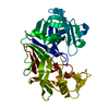
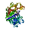
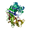
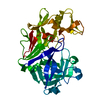
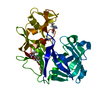
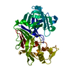
| Entry | Database: PDB / ID: 6scv | ||||||
|---|---|---|---|---|---|---|---|
| Title | Endothiapepsin in complex with ligand 69 | ||||||
 Components Components | Endothiapepsin | ||||||
 Keywords Keywords | HYDROLASE / Aspartic Proteinase / Endothiapepsin / Complex with tetrazole / Inhibitor | ||||||
| Function / homology |  Function and homology information Function and homology information | ||||||
| Biological species |  Cryphonectria parasitica (chestnut blight fungus) Cryphonectria parasitica (chestnut blight fungus) | ||||||
| Method |  X-RAY DIFFRACTION / MOLECULAR REPLACEMENT / Resolution: 1.7 Å X-RAY DIFFRACTION / MOLECULAR REPLACEMENT / Resolution: 1.7 Å | ||||||
 Authors Authors | Magari, F. / Heine, A. / Klebe, G. | ||||||
 Citation Citation | Journal: To Be Published Title: Endothiapepsin in complex with ligand 69 Authors: Magari, F. / Heine, A. / Konstantinidou, M. / Sutanto, F. / Haupenthal, J. / Jumde, R.V. / Unver, M.Y. / Camacho, C.J. / Hirsch, A.K.H. / Doemling, A. / Klebe, G. | ||||||
| History |
|
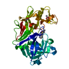
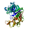
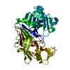
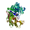
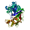
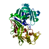
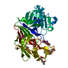
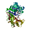
- Structure visualization
| Structure viewer | Molecule: MolmilJmol/JSmol |
|---|
- Downloads & links
-Download
| PDBx/mmCIF format | 6scv.cif.gz | 220 KB | Display | PDBx/mmCIF format |
|---|---|---|---|---|
| PDB format | pdb6scv.ent.gz | 145 KB | Display | PDB format |
| PDBx/mmJSON format | 6scv.json.gz | Tree view | PDBx/mmJSON format | |
| Others |  Other downloads Other downloads |
-Validation report
| Arichive directory | https://data.pdbj.org/pub/pdb/validation_reports/sc/6scvftp://data.pdbj.org/pub/pdb/validation_reports/sc/6scv | HTTPS FTP |
|---|
-Related structure data
| Related structure data | |
|---|---|
| Similar structure data |


-Links
 PDBj
PDBj
- Assembly
| Deposited unit | 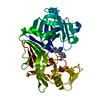
| ||||||||||||
|---|---|---|---|---|---|---|---|---|---|---|---|---|---|
| 1 |
| ||||||||||||
| Unit cell |
|
-Components
| #1: Protein | Mass: 33813.855 Da / Num. of mol.: 1 / Source method: isolated from a natural source Source: (natural) Cryphonectria parasitica (chestnut blight fungus)References: UniProt: P11838, endothiapepsin |
|---|---|
| #2: Chemical | ChemComp-GOL /   Mass: 92.094 Da / Num. of mol.: 1 / Source method: obtained synthetically / Formula: C3H8O3 Mass: 92.094 Da / Num. of mol.: 1 / Source method: obtained synthetically / Formula: C3H8O3 |
| #3: Chemical | ChemComp-L7K / [(~{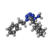  Mass: 300.402 Da / Num. of mol.: 1 / Source method: obtained synthetically / Formula: C16H24N6 / Feature type: SUBJECT OF INVESTIGATION Mass: 300.402 Da / Num. of mol.: 1 / Source method: obtained synthetically / Formula: C16H24N6 / Feature type: SUBJECT OF INVESTIGATION |
| #4: Water | ChemComp-HOH /  Mass: 18.015 Da / Num. of mol.: 274 / Source method: isolated from a natural source / Formula: H2O Mass: 18.015 Da / Num. of mol.: 274 / Source method: isolated from a natural source / Formula: H2O |
| Has ligand of interest | Y |
| Has protein modification | Y |
-Experimental details
-Experiment
| Experiment | Method: X-RAY DIFFRACTION / Number of used crystals: 1 |
|---|
- Sample preparation
| Crystal | Density Matthews: 2.44 Å3/Da / Density % sol: 49.58 % |
|---|---|
| Crystal grow | Temperature: 290 K / Method: vapor diffusion, sitting drop / pH: 4.6 Details: 0.1 M AMMONIUM ACETATE, 0.1M SODIUM ACETATE, 24-30% PEG 4000 CRYSTAL OBTAINED BY STREAK-SEEDING |
-Data collection
| Diffraction | Mean temperature: 113 K / Serial crystal experiment: N |
|---|---|
| Diffraction source | Source: SEALED TUBE / Type: BRUKER IMUS MICROFOCUS / Wavelength: 1.542 Å |
| Detector | Type: MAR scanner 345 mm plate / Detector: IMAGE PLATE / Date: May 31, 2016 |
| Radiation | Protocol: SINGLE WAVELENGTH / Monochromatic (M) / Laue (L): M / Scattering type: x-ray |
| Radiation wavelength | Wavelength: 1.542 Å / Relative weight: 1 |
| Reflection | Resolution: 1.7→50 Å / Num. obs: 35726 / % possible obs: 99.3 % / Redundancy: 4.4 % / Biso Wilson estimate: 13.9 Å2 / CC1/2: 0.999 / Rsym value: 0.038 / Net I/σ(I): 24.79 |
| Reflection shell | Resolution: 1.7→1.8 Å / Mean I/σ(I) obs: 6.26 / Num. unique obs: 5629 / CC1/2: 0.947 / Rsym value: 0.224 / % possible all: 97.7 |
- Processing
| Software |
| ||||||||||||||||||||||||||||||||||||||||||||||||||||||||||||||||||||||||||||||||||||||||||||||||||
|---|---|---|---|---|---|---|---|---|---|---|---|---|---|---|---|---|---|---|---|---|---|---|---|---|---|---|---|---|---|---|---|---|---|---|---|---|---|---|---|---|---|---|---|---|---|---|---|---|---|---|---|---|---|---|---|---|---|---|---|---|---|---|---|---|---|---|---|---|---|---|---|---|---|---|---|---|---|---|---|---|---|---|---|---|---|---|---|---|---|---|---|---|---|---|---|---|---|---|---|
| Refinement | Method to determine structure: MOLECULAR REPLACEMENT Starting model: NONE Resolution: 1.7→39.58 Å / SU ML: 0.1262 / Cross valid method: FREE R-VALUE / σ(F): 1.37 / Phase error: 14.8521
| ||||||||||||||||||||||||||||||||||||||||||||||||||||||||||||||||||||||||||||||||||||||||||||||||||
| Solvent computation | Shrinkage radii: 0.9 Å / VDW probe radii: 1.11 Å | ||||||||||||||||||||||||||||||||||||||||||||||||||||||||||||||||||||||||||||||||||||||||||||||||||
| Displacement parameters | Biso mean: 16.24 Å2 | ||||||||||||||||||||||||||||||||||||||||||||||||||||||||||||||||||||||||||||||||||||||||||||||||||
| Refinement step | Cycle: LAST / Resolution: 1.7→39.58 Å
| ||||||||||||||||||||||||||||||||||||||||||||||||||||||||||||||||||||||||||||||||||||||||||||||||||
| Refine LS restraints |
| ||||||||||||||||||||||||||||||||||||||||||||||||||||||||||||||||||||||||||||||||||||||||||||||||||
| LS refinement shell |
| ||||||||||||||||||||||||||||||||||||||||||||||||||||||||||||||||||||||||||||||||||||||||||||||||||
| Refinement TLS params. | Method: refined / Refine-ID: X-RAY DIFFRACTION
| ||||||||||||||||||||||||||||||||||||||||||||||||||||||||||||||||||||||||||||||||||||||||||||||||||
| Refinement TLS group |
|
