 Movie
Movie Controller

[English] 日本語
 Yorodumi
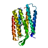
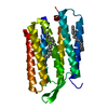
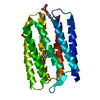
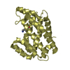
- PDB-6scp: Cell Division Protein SepF in complex with C-terminal domain of FtsZ -
+ Open data

- Basic information
| Entry | Database: PDB / ID: 6scp | ||||||
|---|---|---|---|---|---|---|---|
| Title | Cell Division Protein SepF in complex with C-terminal domain of FtsZ | ||||||
 Components Components | Cell division protein SepF | ||||||
 Keywords Keywords | CELL CYCLE / Cell Division protein | ||||||
| Function / homology |  Function and homology information Function and homology informationdivision septum assembly / FtsZ-dependent cytokinesis / metal ion binding / cytoplasm Similarity search - Function | ||||||
| Biological species |  Corynebacterium glutamicum ATCC 13032 (bacteria) Corynebacterium glutamicum ATCC 13032 (bacteria) | ||||||
| Method |  X-RAY DIFFRACTION / SYNCHROTRON / MOLECULAR REPLACEMENT / molecular replacement / Resolution: 1.8 Å X-RAY DIFFRACTION / SYNCHROTRON / MOLECULAR REPLACEMENT / molecular replacement / Resolution: 1.8 Å | ||||||
 Authors Authors | Sogues, A. / Wehenkel, A.M. / Alzari, P.M. | ||||||
| Funding support |  France, 1items France, 1items
| ||||||
 Citation Citation | Journal: Nat Commun / Year: 2020 Title: Essential dynamic interdependence of FtsZ and SepF for Z-ring and septum formation in Corynebacterium glutamicum. Authors: Sogues, A. / Martinez, M. / Gaday, Q. / Ben Assaya, M. / Grana, M. / Voegele, A. / VanNieuwenhze, M. / England, P. / Haouz, A. / Chenal, A. / Trepout, S. / Duran, R. / Wehenkel, A.M. / Alzari, P.M. | ||||||
| History |
|
- Structure visualization
| Structure viewer | Molecule: MolmilJmol/JSmol |
|---|
- Downloads & links
-Download
| PDBx/mmCIF format | 6scp.cif.gz | 84.5 KB | Display | PDBx/mmCIF format |
|---|---|---|---|---|
| PDB format | pdb6scp.ent.gz | 62.8 KB | Display | PDB format |
| PDBx/mmJSON format | 6scp.json.gz | Tree view | PDBx/mmJSON format | |
| Others |  Other downloads Other downloads |
-Validation report
| Arichive directory | https://data.pdbj.org/pub/pdb/validation_reports/sc/6scpftp://data.pdbj.org/pub/pdb/validation_reports/sc/6scp | HTTPS FTP |
|---|
-Related structure data
| Related structure data |  6satSC  6scqC  6scsC S: Starting model for refinement C: citing same article ( |
|---|---|
| Similar structure data |
-Links
 PDBj
PDBj- Assembly
| Deposited unit | 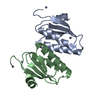
| ||||||||
|---|---|---|---|---|---|---|---|---|---|
| 1 |
| ||||||||
| Unit cell |
|
-Components
| #1: Protein | Mass: 9948.335 Da / Num. of mol.: 2 Source method: isolated from a genetically manipulated source Source: (gene. exp.) Corynebacterium glutamicum ATCC 13032 (bacteria)Gene: sepF, Cgl2152 Production host: References: UniProt: Q8NNN6 #2: Chemical | ChemComp-ZN /   Mass: 65.409 Da / Num. of mol.: 6 / Source method: obtained synthetically / Formula: Zn Mass: 65.409 Da / Num. of mol.: 6 / Source method: obtained synthetically / Formula: Zn#3: Water | ChemComp-HOH / |  Mass: 18.015 Da / Num. of mol.: 109 / Source method: isolated from a natural source / Formula: H2O Mass: 18.015 Da / Num. of mol.: 109 / Source method: isolated from a natural source / Formula: H2OHas ligand of interest | N | |
|---|
-Experimental details
-Experiment
| Experiment | Method: X-RAY DIFFRACTION / Number of used crystals: 1 |
|---|
- Sample preparation
| Crystal | Density Matthews: 2.35 Å3/Da / Density % sol: 47.75 % |
|---|---|
| Crystal grow | Temperature: 291 K / Method: vapor diffusion, sitting drop Details: 10% PEG800, 0.2 M Zinc acetate, 0.1 M sodium acetate. |
-Data collection
| Diffraction | Mean temperature: 100 K / Serial crystal experiment: N |
|---|---|
| Diffraction source | Source: SYNCHROTRON / Site: ESRF / Beamline: ID30B / Wavelength: 0.97625 Å |
| Detector | Type: DECTRIS PILATUS3 6M / Detector: PIXEL / Date: Feb 15, 2018 |
| Radiation | Protocol: SINGLE WAVELENGTH / Monochromatic (M) / Laue (L): M / Scattering type: x-ray |
| Radiation wavelength | Wavelength: 0.97625 Å / Relative weight: 1 |
| Reflection | Resolution: 1.8→46.4 Å / Num. obs: 17334 / % possible obs: 99.9 % / Redundancy: 5.2 % / Biso Wilson estimate: 22.53 Å2 / CC1/2: 0.999 / Rmerge(I) obs: 0.074 / Net I/σ(I): 12.9 |
| Reflection shell | Resolution: 1.8→1.84 Å / Redundancy: 5.2 % / Rmerge(I) obs: 0.551 / Mean I/σ(I) obs: 2.5 / Num. unique obs: 1022 / CC1/2: 0.903 / % possible all: 100 |
-Phasing
| Phasing | Method: molecular replacement |
|---|
- Processing
| Software |
| ||||||||||||||||||||||||||||||||||||||||||||||||||||||||||||||||||||||||||||||||||||||||||||||||||||||||||||
|---|---|---|---|---|---|---|---|---|---|---|---|---|---|---|---|---|---|---|---|---|---|---|---|---|---|---|---|---|---|---|---|---|---|---|---|---|---|---|---|---|---|---|---|---|---|---|---|---|---|---|---|---|---|---|---|---|---|---|---|---|---|---|---|---|---|---|---|---|---|---|---|---|---|---|---|---|---|---|---|---|---|---|---|---|---|---|---|---|---|---|---|---|---|---|---|---|---|---|---|---|---|---|---|---|---|---|---|---|---|
| Refinement | Method to determine structure: MOLECULAR REPLACEMENT Starting model: 6sat Resolution: 1.8→35.53 Å / Cor.coef. Fo:Fc: 0.931 / Cor.coef. Fo:Fc free: 0.929 / SU R Cruickshank DPI: 0.122 / Cross valid method: THROUGHOUT / σ(F): 0 / SU R Blow DPI: 0.129 / SU Rfree Blow DPI: 0.116 / SU Rfree Cruickshank DPI: 0.112
| ||||||||||||||||||||||||||||||||||||||||||||||||||||||||||||||||||||||||||||||||||||||||||||||||||||||||||||
| Displacement parameters | Biso max: 111.59 Å2 / Biso mean: 32.2 Å2 / Biso min: 15.9 Å2
| ||||||||||||||||||||||||||||||||||||||||||||||||||||||||||||||||||||||||||||||||||||||||||||||||||||||||||||
| Refine analyze | Luzzati coordinate error obs: 0.24 Å | ||||||||||||||||||||||||||||||||||||||||||||||||||||||||||||||||||||||||||||||||||||||||||||||||||||||||||||
| Refinement step | Cycle: final / Resolution: 1.8→35.53 Å
| ||||||||||||||||||||||||||||||||||||||||||||||||||||||||||||||||||||||||||||||||||||||||||||||||||||||||||||
| Refine LS restraints |
| ||||||||||||||||||||||||||||||||||||||||||||||||||||||||||||||||||||||||||||||||||||||||||||||||||||||||||||
| LS refinement shell | Resolution: 1.8→1.81 Å / Rfactor Rfree error: 0 / Total num. of bins used: 43
| ||||||||||||||||||||||||||||||||||||||||||||||||||||||||||||||||||||||||||||||||||||||||||||||||||||||||||||
| Refinement TLS params. | Method: refined / Refine-ID: X-RAY DIFFRACTION
| ||||||||||||||||||||||||||||||||||||||||||||||||||||||||||||||||||||||||||||||||||||||||||||||||||||||||||||
| Refinement TLS group |
|
