 Movie
Movie Controller

[English] 日本語
 Yorodumi
- PDB-6s9w: Crystal Structure of AKT1 in Complex with Covalent-Allosteric AKT... -
+ Open data

- Basic information
| Entry | Database: PDB / ID: 6s9w | ||||||||||||
|---|---|---|---|---|---|---|---|---|---|---|---|---|---|
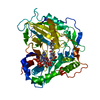
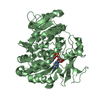
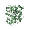
| Title | Crystal Structure of AKT1 in Complex with Covalent-Allosteric AKT Inhibitor 16a | ||||||||||||
 Components Components | RAC-alpha serine/threonine-protein kinase | ||||||||||||
 Keywords Keywords | TRANSFERASE / Akt1 / Akt2 / Akt3 / borussertib / covalent-allosteric | ||||||||||||
| Function / homology |  Function and homology information Function and homology informationpositive regulation of endodeoxyribonuclease activity / regulation of tRNA methylation / negative regulation of protein maturation / negative regulation of fatty acid beta-oxidation / positive regulation of protein localization to endoplasmic reticulum / regulation of glycogen biosynthetic process / negative regulation of lymphocyte migration / cellular response to decreased oxygen levels / cellular response to rapamycin / negative regulation of protein localization to lysosome ...positive regulation of endodeoxyribonuclease activity / regulation of tRNA methylation / negative regulation of protein maturation / negative regulation of fatty acid beta-oxidation / positive regulation of protein localization to endoplasmic reticulum / regulation of glycogen biosynthetic process / negative regulation of lymphocyte migration / cellular response to decreased oxygen levels / cellular response to rapamycin / negative regulation of protein localization to lysosome / maternal placenta development / maintenance of protein location in mitochondrion / mammalian oogenesis stage / AKT-mediated inactivation of FOXO1A / negative regulation of long-chain fatty acid import across plasma membrane / Negative regulation of the PI3K/AKT network / regulation of type B pancreatic cell development / positive regulation of anaphase-promoting complex-dependent catabolic process / : / potassium channel activator activity / sperm glycocalyx / AKT phosphorylates targets in the nucleus / activation-induced cell death of T cells / mammary gland epithelial cell differentiation / cellular response to oxidised low-density lipoprotein particle stimulus / negative regulation of cilium assembly / negative regulation of hydrogen peroxide-induced neuron intrinsic apoptotic signaling pathway / Butyrate Response Factor 1 (BRF1) binds and destabilizes mRNA / fibroblast migration / positive regulation of TORC2 signaling / positive regulation of glucose metabolic process / perinuclear theca / RUNX2 regulates genes involved in cell migration / cellular response to peptide / beta-arrestin-dependent dopamine receptor signaling pathway / positive regulation of organ growth / interleukin-18-mediated signaling pathway / positive regulation of sodium ion transport / MTOR signalling / response to growth factor / cell migration involved in sprouting angiogenesis / peripheral nervous system myelin maintenance / response to fluid shear stress / cellular response to granulocyte macrophage colony-stimulating factor stimulus / negative regulation of leukocyte cell-cell adhesion / RAB GEFs exchange GTP for GDP on RABs / glycogen biosynthetic process / phosphatidylinositol-3,4-bisphosphate binding / positive regulation of protein localization to cell surface / phosphorylation / complement receptor mediated signaling pathway / sphingosine-1-phosphate receptor signaling pathway / response to growth hormone / AKT phosphorylates targets in the cytosol / anoikis / regulation of postsynapse organization / positive regulation of fibroblast migration / labyrinthine layer blood vessel development / execution phase of apoptosis / regulation of myelination / KSRP (KHSRP) binds and destabilizes mRNA / response to UV-A / Regulation of TP53 Activity through Association with Co-factors / TORC2 complex binding / Mechanical load activates signaling by PIEZO1 and integrins in osteocytes / negative regulation of macroautophagy / Co-inhibition by CTLA4 / negative regulation of release of cytochrome c from mitochondria / negative regulation of cGAS/STING signaling pathway / peptidyl-threonine phosphorylation / cellular response to stress / response to food / regulation of neuron projection development / negative regulation of PERK-mediated unfolded protein response / Constitutive Signaling by AKT1 E17K in Cancer / apoptotic mitochondrial changes / positive regulation of protein metabolic process / phosphatidylinositol-3,4,5-trisphosphate binding / Regulation of localization of FOXO transcription factors / negative regulation of Notch signaling pathway / TOR signaling / cellular response to vascular endothelial growth factor stimulus / behavioral response to pain / CD28 dependent PI3K/Akt signaling / positive regulation of peptidyl-serine phosphorylation / Activation of BAD and translocation to mitochondria / positive regulation of blood vessel endothelial cell migration / Estrogen-dependent nuclear events downstream of ESR-membrane signaling / positive regulation of glycogen biosynthetic process / negative regulation of extrinsic apoptotic signaling pathway in absence of ligand / Mitochondrial unfolded protein response (UPRmt) / protein serine/threonine kinase inhibitor activity / response to insulin-like growth factor stimulus / SARS-CoV-2 targets host intracellular signalling and regulatory pathways / positive regulation of lipid biosynthetic process / positive regulation of fat cell differentiation / negative regulation of oxidative stress-induced intrinsic apoptotic signaling pathway / Cyclin E associated events during G1/S transition / positive regulation of G1/S transition of mitotic cell cycle / vascular endothelial cell response to laminar fluid shear stress Similarity search - Function | ||||||||||||
| Biological species |  Homo sapiens (human) Homo sapiens (human) | ||||||||||||
| Method |  X-RAY DIFFRACTION / SYNCHROTRON / MOLECULAR REPLACEMENT / Resolution: 2.3 Å X-RAY DIFFRACTION / SYNCHROTRON / MOLECULAR REPLACEMENT / Resolution: 2.3 Å | ||||||||||||
 Authors Authors | Landel, I. / Mueller, M.P. / Rauh, D. | ||||||||||||
| Funding support |  Germany, 3items Germany, 3items
| ||||||||||||
 Citation Citation | Journal: Angew.Chem.Int.Ed.Engl. / Year: 2019 Title: Covalent-Allosteric Inhibitors to Achieve Akt Isoform-Selectivity. Authors: Quambusch, L. / Landel, I. / Depta, L. / Weisner, J. / Uhlenbrock, N. / Muller, M.P. / Glanemann, F. / Althoff, K. / Siveke, J.T. / Rauh, D. | ||||||||||||
| History |
|
- Structure visualization
| Structure viewer | Molecule: MolmilJmol/JSmol |
|---|
- Downloads & links
-Download
| PDBx/mmCIF format | 6s9w.cif.gz | 186.4 KB | Display | PDBx/mmCIF format |
|---|---|---|---|---|
| PDB format | pdb6s9w.ent.gz | 147.3 KB | Display | PDB format |
| PDBx/mmJSON format | 6s9w.json.gz | Tree view | PDBx/mmJSON format | |
| Others |  Other downloads Other downloads |
-Validation report
| Arichive directory | https://data.pdbj.org/pub/pdb/validation_reports/s9/6s9wftp://data.pdbj.org/pub/pdb/validation_reports/s9/6s9w | HTTPS FTP |
|---|
-Related structure data
| Related structure data |  6s9xC  6hhgS S: Starting model for refinement C: citing same article ( |
|---|---|
| Similar structure data |


-Links
 PDBj
PDBj


- Assembly
| Deposited unit | 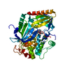
| ||||||||
|---|---|---|---|---|---|---|---|---|---|
| 1 |
| ||||||||
| Unit cell |
|
-Components
| #1: Protein | Mass: 51746.035 Da / Num. of mol.: 1 Source method: isolated from a genetically manipulated source Source: (gene. exp.) Homo sapiens (human) / Gene: AKT1, PKB, RAC / Production host:   Spodoptera frugiperda (fall armyworm) Spodoptera frugiperda (fall armyworm)References: UniProt: P31749, non-specific serine/threonine protein kinase |
|---|---|
| #2: Chemical | ChemComp-L1Z / ~{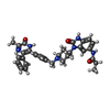  Mass: 562.661 Da / Num. of mol.: 1 / Source method: obtained synthetically / Formula: C33H34N6O3 / Feature type: SUBJECT OF INVESTIGATION Mass: 562.661 Da / Num. of mol.: 1 / Source method: obtained synthetically / Formula: C33H34N6O3 / Feature type: SUBJECT OF INVESTIGATION |
| #3: Water | ChemComp-HOH /  Mass: 18.015 Da / Num. of mol.: 12 / Source method: isolated from a natural source / Formula: H2O Mass: 18.015 Da / Num. of mol.: 12 / Source method: isolated from a natural source / Formula: H2O |
| Has ligand of interest | Y |
| Has protein modification | N |
-Experimental details
-Experiment
| Experiment | Method: X-RAY DIFFRACTION / Number of used crystals: 1 |
|---|
- Sample preparation
| Crystal | Density Matthews: 2.2 Å3/Da / Density % sol: 44.08 % |
|---|---|
| Crystal grow | Temperature: 293 K / Method: vapor diffusion, hanging drop / pH: 7 Details: 1.25 mM Na-acetate, 3.75 mM Na-citrate, 24% v/v PEG 2000 MME, pH 7.0, 3 mg/mL Akt1 (in 25 mM TRIS, 100 mM NaCl, 10% v/v Glycerol, 5 mM DTT, pH 7.5), 1 uL reservoir + 1 uL protein solution |
-Data collection
| Diffraction | Mean temperature: 100 K / Serial crystal experiment: N |
|---|---|
| Diffraction source | Source: SYNCHROTRON / Site: SLS  / Beamline: X10SA / Wavelength: 0.91886 Å / Beamline: X10SA / Wavelength: 0.91886 Å |
| Detector | Type: DECTRIS PILATUS 6M / Detector: PIXEL / Date: May 20, 2018 |
| Radiation | Monochromator: Si(111) / Protocol: SINGLE WAVELENGTH / Monochromatic (M) / Laue (L): M / Scattering type: x-ray |
| Radiation wavelength | Wavelength: 0.91886 Å / Relative weight: 1 |
| Reflection | Resolution: 2.3→45.67 Å / Num. obs: 19153 / % possible obs: 91.8 % / Redundancy: 12.7 % / CC1/2: 0.999 / Rmerge(I) obs: 0.098 / Rrim(I) all: 0.102 / Net I/σ(I): 15.06 |
| Reflection shell | Resolution: 2.3→2.4 Å / Redundancy: 12.9 % / Rmerge(I) obs: 1.637 / Mean I/σ(I) obs: 1.62 / Num. unique obs: 2451 / CC1/2: 0.629 / Rrim(I) all: 1.705 / % possible all: 100 |
- Processing
| Software |
| ||||||||||||||||||||||||||||||||||||||||||||||||
|---|---|---|---|---|---|---|---|---|---|---|---|---|---|---|---|---|---|---|---|---|---|---|---|---|---|---|---|---|---|---|---|---|---|---|---|---|---|---|---|---|---|---|---|---|---|---|---|---|---|
| Refinement | Method to determine structure: MOLECULAR REPLACEMENT Starting model: 6HHG Resolution: 2.3→45.67 Å / SU ML: 0.35 / Cross valid method: THROUGHOUT / σ(F): 1.36 / Phase error: 30.82
| ||||||||||||||||||||||||||||||||||||||||||||||||
| Solvent computation | Shrinkage radii: 0.9 Å / VDW probe radii: 1.11 Å | ||||||||||||||||||||||||||||||||||||||||||||||||
| Displacement parameters | Biso max: 167.36 Å2 / Biso mean: 70.9623 Å2 / Biso min: 28.85 Å2 | ||||||||||||||||||||||||||||||||||||||||||||||||
| Refinement step | Cycle: final / Resolution: 2.3→45.67 Å
| ||||||||||||||||||||||||||||||||||||||||||||||||
| LS refinement shell | Refine-ID: X-RAY DIFFRACTION / Rfactor Rfree error: 0
| ||||||||||||||||||||||||||||||||||||||||||||||||
| Refinement TLS params. | Method: refined / Origin x: -13.6844 Å / Origin y: -12.4833 Å / Origin z: 15.0602 Å
| ||||||||||||||||||||||||||||||||||||||||||||||||
| Refinement TLS group | Selection details: (chain A) |
