 Movie
Movie Controller

[English] 日本語
 Yorodumi
- PDB-6s99: Dimethylated fusion protein of RSL and trimeric coiled coil in co... -
+ Open data

- Basic information
| Entry | Database: PDB / ID: 6s99 | |||||||||
|---|---|---|---|---|---|---|---|---|---|---|
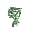
| Title | Dimethylated fusion protein of RSL and trimeric coiled coil in complex with cucurbit[7]uril | |||||||||
 Components Components | 4dzn-RSL,Fucose-binding lectin protein,Fucose-binding lectin protein | |||||||||
 Keywords Keywords | SUGAR BINDING PROTEIN / fusion protein / molecular glues / crystal engineering / cucurbituril | |||||||||
| Function / homology | Fucose-specific lectin / Fungal fucose-specific lectin / carbohydrate binding / metal ion binding / beta-D-mannopyranose / cucurbit[7]uril / Fucose-binding lectin protein Function and homology information Function and homology information | |||||||||
| Biological species |  Ralstonia solanacearum (bacteria) Ralstonia solanacearum (bacteria) | |||||||||
| Method |  X-RAY DIFFRACTION / SYNCHROTRON / MOLECULAR REPLACEMENT / molecular replacement / Resolution: 2.649 Å X-RAY DIFFRACTION / SYNCHROTRON / MOLECULAR REPLACEMENT / molecular replacement / Resolution: 2.649 Å | |||||||||
 Authors Authors | Ramberg, K. / Engilberge, S. / Crowley, P.B. | |||||||||
| Funding support |  Ireland, 1items Ireland, 1items
| |||||||||
 Citation Citation | Journal: Chemistry / Year: 2021 Title: Segregated Protein-Cucurbit[7]uril Crystalline Architectures via Modulatory Peptide Tectons. Authors: Ramberg, K.O. / Guagnini, F. / Engilberge, S. / Wronska, M.A. / Rennie, M.L. / Perez, J. / Crowley, P.B. | |||||||||
| History |
|
- Structure visualization
| Structure viewer | Molecule: MolmilJmol/JSmol |
|---|
- Downloads & links
-Download
| PDBx/mmCIF format | 6s99.cif.gz | 292.7 KB | Display | PDBx/mmCIF format |
|---|---|---|---|---|
| PDB format | pdb6s99.ent.gz | 245.3 KB | Display | PDB format |
| PDBx/mmJSON format | 6s99.json.gz | Tree view | PDBx/mmJSON format | |
| Others |  Other downloads Other downloads |
-Validation report
| Arichive directory | https://data.pdbj.org/pub/pdb/validation_reports/s9/6s99ftp://data.pdbj.org/pub/pdb/validation_reports/s9/6s99 | HTTPS FTP |
|---|
-Related structure data


-Links
 PDBj
PDBj
- Assembly
| Deposited unit | 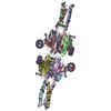
| ||||||||
|---|---|---|---|---|---|---|---|---|---|
| 1 | 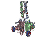
| ||||||||
| 2 | 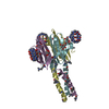
| ||||||||
| Unit cell |
|
-Components
| #1: Protein | Mass: 12374.828 Da / Num. of mol.: 6 Source method: isolated from a genetically manipulated source Source: (gene. exp.) Ralstonia solanacearum (bacteria)Gene: E7Z57_08365, HF909_06975, HXP36_18875, RSP795_21825, RSP822_19650, RUN39_v1_50103 Production host: #2: Sugar | ChemComp-BMA /   Type: D-saccharide, beta linking / Mass: 180.156 Da / Num. of mol.: 12 / Source method: obtained synthetically / Formula: C6H12O6 Type: D-saccharide, beta linking / Mass: 180.156 Da / Num. of mol.: 12 / Source method: obtained synthetically / Formula: C6H12O6#3: Chemical | ChemComp-QQ7 /   Mass: 1162.962 Da / Num. of mol.: 6 / Source method: obtained synthetically / Formula: C42H42N28O14 / Feature type: SUBJECT OF INVESTIGATION Mass: 1162.962 Da / Num. of mol.: 6 / Source method: obtained synthetically / Formula: C42H42N28O14 / Feature type: SUBJECT OF INVESTIGATION#4: Water | ChemComp-HOH / |  Mass: 18.015 Da / Num. of mol.: 112 / Source method: isolated from a natural source / Formula: H2O Mass: 18.015 Da / Num. of mol.: 112 / Source method: isolated from a natural source / Formula: H2OHas ligand of interest | Y | |
|---|
-Experimental details
-Experiment
| Experiment | Method: X-RAY DIFFRACTION / Number of used crystals: 1 |
|---|
- Sample preparation
| Crystal | Density Matthews: 3.35 Å3/Da / Density % sol: 63.26 % |
|---|---|
| Crystal grow | Temperature: 293 K / Method: vapor diffusion, hanging drop Details: 0.1M TRIS HCL pH 8.5, 20% PEG8000, 0.2 M Magnesium Chloride, 0.0013 M cucurbit[7]uril |
-Data collection
| Diffraction | Mean temperature: 100 K / Serial crystal experiment: N |
|---|---|
| Diffraction source | Source: SYNCHROTRON / Site: SOLEIL  / Beamline: PROXIMA 2 / Wavelength: 0.98 Å / Beamline: PROXIMA 2 / Wavelength: 0.98 Å |
| Detector | Type: DECTRIS EIGER X 9M / Detector: PIXEL / Date: Sep 21, 2018 |
| Radiation | Protocol: SINGLE WAVELENGTH / Monochromatic (M) / Laue (L): M / Scattering type: x-ray |
| Radiation wavelength | Wavelength: 0.98 Å / Relative weight: 1 |
| Reflection | Resolution: 2.649→42.842 Å / Num. obs: 26787 / % possible obs: 97.4 % / Redundancy: 3.6 % / Rmerge(I) obs: 0.056 / Rpim(I) all: 0.035 / Net I/σ(I): 7.8 |
| Reflection shell | Resolution: 2.649→2.695 Å / Rmerge(I) obs: 0.313 / Mean I/σ(I) obs: 2.7 / Num. unique obs: 1383 / Rpim(I) all: 0.19 |
-Phasing
| Phasing | Method: molecular replacement |
|---|
- Processing
| Software |
| |||||||||||||||||||||||||||||||||||||||||||||||||||||||||||||||||||||||||||||
|---|---|---|---|---|---|---|---|---|---|---|---|---|---|---|---|---|---|---|---|---|---|---|---|---|---|---|---|---|---|---|---|---|---|---|---|---|---|---|---|---|---|---|---|---|---|---|---|---|---|---|---|---|---|---|---|---|---|---|---|---|---|---|---|---|---|---|---|---|---|---|---|---|---|---|---|---|---|---|
| Refinement | Method to determine structure: MOLECULAR REPLACEMENT / Resolution: 2.649→42.842 Å / SU ML: 0.38 / Cross valid method: FREE R-VALUE / σ(F): 2.1 / Phase error: 30.06
| |||||||||||||||||||||||||||||||||||||||||||||||||||||||||||||||||||||||||||||
| Solvent computation | Shrinkage radii: 0.9 Å / VDW probe radii: 1.11 Å | |||||||||||||||||||||||||||||||||||||||||||||||||||||||||||||||||||||||||||||
| Refinement step | Cycle: LAST / Resolution: 2.649→42.842 Å
| |||||||||||||||||||||||||||||||||||||||||||||||||||||||||||||||||||||||||||||
| Refine LS restraints |
| |||||||||||||||||||||||||||||||||||||||||||||||||||||||||||||||||||||||||||||
| LS refinement shell |
| |||||||||||||||||||||||||||||||||||||||||||||||||||||||||||||||||||||||||||||
| Refinement TLS params. | Method: refined / Origin x: -0.7799 Å / Origin y: 0.1014 Å / Origin z: -1.2297 Å
| |||||||||||||||||||||||||||||||||||||||||||||||||||||||||||||||||||||||||||||
| Refinement TLS group | Selection details: all |
