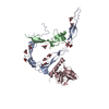
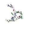
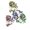
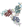
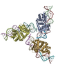
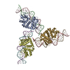
Crystal Structure of Properdin (TSR domains N12 & 456)
Components
(Properdin) x 2
Keywords
IMMUNE SYSTEM / INNATE IMMUNITY / COMPLEMENT
Function / homology
Function and homology information
positive regulation of complement activation, alternative pathway / cytoplasmic side of Golgi membrane / Defective B3GALTL causes PpS / O-glycosylation of TSR domain-containing proteins / positive regulation of opsonization / Alternative complement activation / Activation of C3 and C5 / complement activation / complement activation, alternative pathway / Regulation of Complement cascade ...positive regulation of complement activation, alternative pathway / cytoplasmic side of Golgi membrane / Defective B3GALTL causes PpS / O-glycosylation of TSR domain-containing proteins / positive regulation of opsonization / Alternative complement activation / Activation of C3 and C5 / complement activation / complement activation, alternative pathway / Regulation of Complement cascade / specific granule lumen / positive regulation of immune response / tertiary granule lumen / defense response to bacterium / protein stabilization / immune response / endoplasmic reticulum lumen / Neutrophil degranulation / : / extracellular region Similarity search - Function
Method to determine structure: MOLECULAR REPLACEMENT / Resolution: 2.52→102.881 Å / Cor.coef. Fo:Fc: 0.893 / Cor.coef. Fo:Fc free: 0.885 / SU B: 27.828 / SU ML: 0.256 / Cross valid method: THROUGHOUT / σ(F): 0 / ESU R: 0.654 / ESU R Free: 0.35 Details: U VALUES : WITH TLS ADDED HYDROGENS HAVE BEEN ADDED IN THE RIDING POSITIONS
Rfactor
Num. reflection
% reflection
Selection details
Rfree
0.2672
770
4.7 %
RANDOM
Rwork
0.248
-
-
-
obs
0.2489
15443
60.47 %
-
Solvent computation
Ion probe radii: 0.8 Å / Shrinkage radii: 0.8 Å / VDW probe radii: 1.2 Å
In the structure databanks used in Yorodumi, some data are registered as the other names, "COVID-19 virus" and "2019-nCoV". Here are the details of the virus and the list of structure data.
Jan 31, 2019. EMDB accession codes are about to change! (news from PDBe EMDB page)
EMDB accession codes are about to change! (news from PDBe EMDB page)
The allocation of 4 digits for EMDB accession codes will soon come to an end. Whilst these codes will remain in use, new EMDB accession codes will include an additional digit and will expand incrementally as the available range of codes is exhausted. The current 4-digit format prefixed with “EMD-” (i.e. EMD-XXXX) will advance to a 5-digit format (i.e. EMD-XXXXX), and so on. It is currently estimated that the 4-digit codes will be depleted around Spring 2019, at which point the 5-digit format will come into force.
The EM Navigator/Yorodumi systems omit the EMD- prefix.
Related info.:Q: What is EMD? / ID/Accession-code notation in Yorodumi/EM Navigator
Yorodumi is a browser for structure data from EMDB, PDB, SASBDB, etc.
This page is also the successor to EM Navigator detail page, and also detail information page/front-end page for Omokage search.
The word "yorodu" (or yorozu) is an old Japanese word meaning "ten thousand". "mi" (miru) is to see.
Related info.:EMDB / PDB / SASBDB / Comparison of 3 databanks / Yorodumi Search / Aug 31, 2016. New EM Navigator & Yorodumi / Yorodumi Papers / Jmol/JSmol / Function and homology information / Changes in new EM Navigator and Yorodumi
 Movie
Movie Controller


 Open data
 Basic information
 Components
 Keywords
 Function and homology information
 Homo sapiens (human)
 X-RAY DIFFRACTION /
  Authors
 Netherlands, 1items
  Citation
 Structure visualization
 Downloads & links
 Other downloads
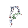
 PDBj
PDBj


 Assembly
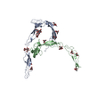


 Type: D-saccharide, alpha linking / Mass: 180.156 Da / Num. of mol.: 10
 Type: L-saccharide, alpha linking / Mass: 164.156 Da / Num. of mol.: 1
 Sample preparation
 / Beamline: ID29 / Wavelength: 0.9763 Å
 Processing