 Movie
Movie Controller


+ Open data

- Basic information
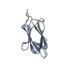
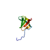
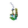
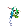
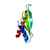
| Entry | Database: PDB / ID: 6ry3 | ||||||
|---|---|---|---|---|---|---|---|
| Title | Structure of the WUS homeodomain | ||||||
 Components Components | Protein WUSCHEL | ||||||
 Keywords Keywords | DNA BINDING PROTEIN / Homeodomain Transcription factor | ||||||
| Function / homology |  Function and homology information Function and homology informationstomium development / anther development / axillary shoot meristem initiation / stem cell population maintenance / cell differentiation / transcription cis-regulatory region binding / DNA-binding transcription factor activity / regulation of DNA-templated transcription / nucleus Similarity search - Function | ||||||
| Biological species |  | ||||||
| Method |  X-RAY DIFFRACTION / SYNCHROTRON / MOLECULAR REPLACEMENT / Resolution: 1.374 Å X-RAY DIFFRACTION / SYNCHROTRON / MOLECULAR REPLACEMENT / Resolution: 1.374 Å | ||||||
 Authors Authors | Sloan, J.J. / Wild, K. / Sinning, I. | ||||||
| Funding support |  Germany, 1items Germany, 1items
| ||||||
 Citation Citation | Journal: Nat Commun / Year: 2020 Title: Structural basis for the complex DNA binding behavior of the plant stem cell regulator WUSCHEL. Authors: Sloan, J. / Hakenjos, J.P. / Gebert, M. / Ermakova, O. / Gumiero, A. / Stier, G. / Wild, K. / Sinning, I. / Lohmann, J.U. | ||||||
| History |
|
- Structure visualization
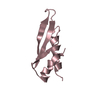
| Structure viewer | Molecule: MolmilJmol/JSmol |
|---|
- Downloads & links
-Download
| PDBx/mmCIF format | 6ry3.cif.gz | 44.7 KB | Display | PDBx/mmCIF format |
|---|---|---|---|---|
| PDB format | pdb6ry3.ent.gz | 29.9 KB | Display | PDB format |
| PDBx/mmJSON format | 6ry3.json.gz | Tree view | PDBx/mmJSON format | |
| Others |  Other downloads Other downloads |
-Validation report
| Arichive directory | https://data.pdbj.org/pub/pdb/validation_reports/ry/6ry3ftp://data.pdbj.org/pub/pdb/validation_reports/ry/6ry3 | HTTPS FTP |
|---|
-Related structure data
| Related structure data |  6rydC  6ryiC  6rylC  3hddS S: Starting model for refinement C: citing same article ( |
|---|---|
| Similar structure data |


-Links
 PDBj
PDBj
- Assembly
| Deposited unit | 
| ||||||||
|---|---|---|---|---|---|---|---|---|---|
| 1 |
| ||||||||
| Unit cell |
|
-Components
| #1: Protein | Mass: 8949.188 Da / Num. of mol.: 1 Source method: isolated from a genetically manipulated source Source: (gene. exp.)  | ||||
|---|---|---|---|---|---|
| #2: Chemical | ChemComp-SO4 /   Mass: 96.063 Da / Num. of mol.: 1 / Source method: obtained synthetically / Formula: SO4 Mass: 96.063 Da / Num. of mol.: 1 / Source method: obtained synthetically / Formula: SO4 | ||||
| #3: Chemical |   Mass: 44.053 Da / Num. of mol.: 3 / Source method: obtained synthetically / Formula: C2H4O Mass: 44.053 Da / Num. of mol.: 3 / Source method: obtained synthetically / Formula: C2H4O#4: Water | ChemComp-HOH / |  Mass: 18.015 Da / Num. of mol.: 50 / Source method: isolated from a natural source / Formula: H2O Mass: 18.015 Da / Num. of mol.: 50 / Source method: isolated from a natural source / Formula: H2OHas ligand of interest | N | |
-Experimental details
-Experiment
| Experiment | Method: X-RAY DIFFRACTION / Number of used crystals: 1 |
|---|
- Sample preparation
| Crystal | Density Matthews: 2.41 Å3/Da / Density % sol: 48.93 % |
|---|---|
| Crystal grow | Temperature: 291 K / Method: vapor diffusion, sitting drop Details: 2.5 M ammonium sulfate 0.05 M MES (pH 6.0) 0.01 M magnesium acetate |
-Data collection
| Diffraction | Mean temperature: 100 K / Serial crystal experiment: N |
|---|---|
| Diffraction source | Source: SYNCHROTRON / Site: ESRF  / Beamline: MASSIF-3 / Wavelength: 0.9677 Å / Beamline: MASSIF-3 / Wavelength: 0.9677 Å |
| Detector | Type: DECTRIS EIGER X 4M / Detector: PIXEL / Date: Sep 25, 2016 |
| Radiation | Protocol: SINGLE WAVELENGTH / Monochromatic (M) / Laue (L): M / Scattering type: x-ray |
| Radiation wavelength | Wavelength: 0.9677 Å / Relative weight: 1 |
| Reflection | Resolution: 1.374→38.17 Å / Num. obs: 16816 / % possible obs: 99.85 % / Redundancy: 24.5 % / Biso Wilson estimate: 13.28 Å2 / CC1/2: 0.999 / Rmerge(I) obs: 0.03774 / Rpim(I) all: 0.00818 / Net I/σ(I): 61.11 |
| Reflection shell | Resolution: 1.374→1.423 Å / Num. unique obs: 1628 / Rpim(I) all: 0.03016 |
- Processing
| Software |
| |||||||||||||||||||||||||||||||||||||||||||||||||
|---|---|---|---|---|---|---|---|---|---|---|---|---|---|---|---|---|---|---|---|---|---|---|---|---|---|---|---|---|---|---|---|---|---|---|---|---|---|---|---|---|---|---|---|---|---|---|---|---|---|---|
| Refinement | Method to determine structure: MOLECULAR REPLACEMENT Starting model: 3HDD Resolution: 1.374→38.166 Å / SU ML: 0.15 / Cross valid method: THROUGHOUT / σ(F): 1.39 / Phase error: 22.31 / Stereochemistry target values: ML
| |||||||||||||||||||||||||||||||||||||||||||||||||
| Solvent computation | Shrinkage radii: 0.9 Å / VDW probe radii: 1.11 Å / Solvent model: FLAT BULK SOLVENT MODEL | |||||||||||||||||||||||||||||||||||||||||||||||||
| Displacement parameters | Biso max: 60.51 Å2 / Biso mean: 19.5167 Å2 / Biso min: 6.64 Å2 | |||||||||||||||||||||||||||||||||||||||||||||||||
| Refinement step | Cycle: final / Resolution: 1.374→38.166 Å
| |||||||||||||||||||||||||||||||||||||||||||||||||
| Refine LS restraints |
| |||||||||||||||||||||||||||||||||||||||||||||||||
| LS refinement shell | Refine-ID: X-RAY DIFFRACTION / Rfactor Rfree error: 0 / Total num. of bins used: 6
|
