 Movie
Movie Controller


+ Open data

- Basic information
| Entry | Database: PDB / ID: 6rwf | |||||||||
|---|---|---|---|---|---|---|---|---|---|---|
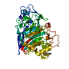
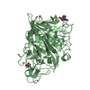
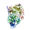
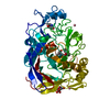
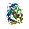
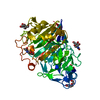
| Title | The dissociation mechanism of processive cellulases | |||||||||
 Components Components | Glucanase | |||||||||
 Keywords Keywords | HYDROLASE / Cellobiogydrolase I / Cel7A from Hypocrea jecorina | |||||||||
| Function / homology |  Function and homology information Function and homology informationHydrolases; Glycosylases; Glycosidases, i.e. enzymes that hydrolyse O- and S-glycosyl compounds / cellulose binding / cellulose catabolic process / hydrolase activity, hydrolyzing O-glycosyl compounds / extracellular region Similarity search - Function | |||||||||
| Biological species |  Hypocrea jecorina (fungus) Hypocrea jecorina (fungus) | |||||||||
| Method |  X-RAY DIFFRACTION / SYNCHROTRON / MOLECULAR REPLACEMENT / Resolution: 1.64 Å X-RAY DIFFRACTION / SYNCHROTRON / MOLECULAR REPLACEMENT / Resolution: 1.64 Å | |||||||||
 Authors Authors | Stahlberg, J. / Knott, B.C. | |||||||||
| Funding support |  United States, 1items United States, 1items
| |||||||||
 Citation Citation | Journal: Proc.Natl.Acad.Sci.USA / Year: 2019 Title: The dissociation mechanism of processive cellulases. Authors: Vermaas, J.V. / Kont, R. / Beckham, G.T. / Crowley, M.F. / Gudmundsson, M. / Sandgren, M. / Stahlberg, J. / Valjamae, P. / Knott, B.C. | |||||||||
| History |
|
- Structure visualization
| Structure viewer | Molecule: MolmilJmol/JSmol |
|---|
- Downloads & links
-Download
| PDBx/mmCIF format | 6rwf.cif.gz | 116.6 KB | Display | PDBx/mmCIF format |
|---|---|---|---|---|
| PDB format | pdb6rwf.ent.gz | 85.2 KB | Display | PDB format |
| PDBx/mmJSON format | 6rwf.json.gz | Tree view | PDBx/mmJSON format | |
| Others |  Other downloads Other downloads |
-Validation report
| Summary document | 6rwf_validation.pdf.gz | 448.5 KB | Display | wwPDB validaton report |
|---|---|---|---|---|
| Full document | 6rwf_full_validation.pdf.gz | 450.1 KB | Display | |
| Data in XML | 6rwf_validation.xml.gz | 24.2 KB | Display | |
| Data in CIF | 6rwf_validation.cif.gz | 38.9 KB | Display | |
| Arichive directory | https://data.pdbj.org/pub/pdb/validation_reports/rw/6rwfftp://data.pdbj.org/pub/pdb/validation_reports/rw/6rwf | HTTPS FTP |
-Related structure data
| Related structure data |  6celS S: Starting model for refinement |
|---|---|
| Similar structure data |


-Links
 PDBj
PDBj
- Assembly
| Deposited unit | 
| ||||||||
|---|---|---|---|---|---|---|---|---|---|
| 1 |
| ||||||||
| Unit cell |
|
-Components
| #1: Protein | Mass: 45808.582 Da / Num. of mol.: 1 Source method: isolated from a genetically manipulated source Source: (gene. exp.) Hypocrea jecorina (strain QM6a) (fungus)Strain: QM6a / Gene: cel7a, TRIREDRAFT_123989 / Production host: Trichoderma reesei QM6a (fungus)References: UniProt: G0RVK1, Hydrolases; Glycosylases; Glycosidases, i.e. enzymes that hydrolyse O- and S-glycosyl compounds | ||||||
|---|---|---|---|---|---|---|---|
| #2: Chemical | ChemComp-CO /   Mass: 58.933 Da / Num. of mol.: 5 / Source method: obtained synthetically / Formula: Co Mass: 58.933 Da / Num. of mol.: 5 / Source method: obtained synthetically / Formula: Co#3: Sugar | ChemComp-NAG / |   Type: D-saccharide, beta linking / Mass: 221.208 Da / Num. of mol.: 1 Type: D-saccharide, beta linking / Mass: 221.208 Da / Num. of mol.: 1Source method: isolated from a genetically manipulated source Formula: C8H15NO6 #4: Water | ChemComp-HOH / |  Mass: 18.015 Da / Num. of mol.: 680 / Source method: isolated from a natural source / Formula: H2O Mass: 18.015 Da / Num. of mol.: 680 / Source method: isolated from a natural source / Formula: H2OHas protein modification | Y | |
-Experimental details
-Experiment
| Experiment | Method: X-RAY DIFFRACTION / Number of used crystals: 1 |
|---|
- Sample preparation
| Crystal | Density Matthews: 2.03 Å3/Da / Density % sol: 39.53 % |
|---|---|
| Crystal grow | Temperature: 293 K / Method: vapor diffusion, hanging drop Details: 10 mM sodium acetate, pH 5.0, 20% PEG 5000 monomethyl ether, 0.1 M MES pH 6.0, 10 mM cobalt chloride, 12.5% glycerol. PH range: 5 - 6 |
-Data collection
| Diffraction | Mean temperature: 100 K / Serial crystal experiment: N |
|---|---|
| Diffraction source | Source: SYNCHROTRON / Site: MAX II  / Beamline: I711 / Wavelength: 0.967 Å / Beamline: I711 / Wavelength: 0.967 Å |
| Detector | Type: MAR CCD 165 mm / Detector: CCD / Date: Sep 6, 2003 |
| Radiation | Protocol: SINGLE WAVELENGTH / Monochromatic (M) / Laue (L): M / Scattering type: x-ray |
| Radiation wavelength | Wavelength: 0.967 Å / Relative weight: 1 |
| Reflection | Resolution: 1.64→40.48 Å / Num. obs: 44198 / % possible obs: 97.5 % / Redundancy: 1.6 % / Net I/σ(I): 8.9 |
| Reflection shell | Resolution: 1.64→1.68 Å / Redundancy: 1.5 % / Num. unique obs: 2624 / % possible all: 91.1 |
- Processing
| Software |
| |||||||||||||||||||||||||||||||||||||||||||||||||||||||||||||||||||||||||||
|---|---|---|---|---|---|---|---|---|---|---|---|---|---|---|---|---|---|---|---|---|---|---|---|---|---|---|---|---|---|---|---|---|---|---|---|---|---|---|---|---|---|---|---|---|---|---|---|---|---|---|---|---|---|---|---|---|---|---|---|---|---|---|---|---|---|---|---|---|---|---|---|---|---|---|---|---|
| Refinement | Method to determine structure: MOLECULAR REPLACEMENT Starting model: 6CEL Resolution: 1.64→29.52 Å / Cor.coef. Fo:Fc: 0.97 / Cor.coef. Fo:Fc free: 0.95 / Cross valid method: THROUGHOUT / σ(F): 0 / ESU R: 0.09 / ESU R Free: 0.09 Details: HYDROGENS HAVE BEEN ADDED IN THE RIDING POSITIONS U VALUES : REFINED INDIVIDUALLY
| |||||||||||||||||||||||||||||||||||||||||||||||||||||||||||||||||||||||||||
| Solvent computation | Ion probe radii: 0.8 Å / Shrinkage radii: 0.8 Å / VDW probe radii: 1.2 Å | |||||||||||||||||||||||||||||||||||||||||||||||||||||||||||||||||||||||||||
| Displacement parameters | Biso max: 90.96 Å2 / Biso mean: 7.707 Å2 / Biso min: 0.95 Å2
| |||||||||||||||||||||||||||||||||||||||||||||||||||||||||||||||||||||||||||
| Refinement step | Cycle: final / Resolution: 1.64→29.52 Å
| |||||||||||||||||||||||||||||||||||||||||||||||||||||||||||||||||||||||||||
| Refine LS restraints |
| |||||||||||||||||||||||||||||||||||||||||||||||||||||||||||||||||||||||||||
| LS refinement shell | Resolution: 1.636→1.678 Å / Total num. of bins used: 20
|
