 Movie
Movie Controller

[English] 日本語
 Yorodumi
- PDB-6rqn: X-ray crystal structure of protiated (H) small monoclinic unit ce... -
+ Open data

- Basic information
| Entry | Database: PDB / ID: 6rqn | ||||||
|---|---|---|---|---|---|---|---|
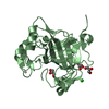
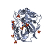
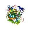
| Title | X-ray crystal structure of protiated (H) small monoclinic unit cell CA IX SV. | ||||||
 Components Components | Carbonic anhydrase 9 | ||||||
 Keywords Keywords | PROTON TRANSPORT / carbonic anhydrase / CA IX / surface variant | ||||||
| Function / homology |  Function and homology information Function and homology informationRegulation of gene expression by Hypoxia-inducible Factor / response to testosterone / microvillus membrane / secretion / Reversible hydration of carbon dioxide / molecular function activator activity / morphogenesis of an epithelium / carbonic anhydrase / carbonate dehydratase activity / basolateral plasma membrane ...Regulation of gene expression by Hypoxia-inducible Factor / response to testosterone / microvillus membrane / secretion / Reversible hydration of carbon dioxide / molecular function activator activity / morphogenesis of an epithelium / carbonic anhydrase / carbonate dehydratase activity / basolateral plasma membrane / response to hypoxia / response to xenobiotic stimulus / nucleolus / zinc ion binding / membrane / plasma membrane Similarity search - Function | ||||||
| Biological species |  Homo sapiens (human) Homo sapiens (human) | ||||||
| Method |  X-RAY DIFFRACTION / SYNCHROTRON / MOLECULAR REPLACEMENT / Resolution: 1.777 Å X-RAY DIFFRACTION / SYNCHROTRON / MOLECULAR REPLACEMENT / Resolution: 1.777 Å | ||||||
 Authors Authors | Fisher, Z. / Koruza, K. | ||||||
 Citation Citation | Journal: Acta Crystallogr D Struct Biol / Year: 2019 Title: Structural comparison of protiated, H/D-exchanged and deuterated human carbonic anhydrase IX. Authors: Koruza, K. / Lafumat, B. / Nyblom, M. / Mahon, B.P. / Knecht, W. / McKenna, R. / Fisher, S.Z. | ||||||
| History |
|
- Structure visualization
| Structure viewer | Molecule: MolmilJmol/JSmol |
|---|
- Downloads & links
-Download
| PDBx/mmCIF format | 6rqn.cif.gz | 74.6 KB | Display | PDBx/mmCIF format |
|---|---|---|---|---|
| PDB format | pdb6rqn.ent.gz | 53.1 KB | Display | PDB format |
| PDBx/mmJSON format | 6rqn.json.gz | Tree view | PDBx/mmJSON format | |
| Others |  Other downloads Other downloads |
-Validation report
| Arichive directory | https://data.pdbj.org/pub/pdb/validation_reports/rq/6rqnftp://data.pdbj.org/pub/pdb/validation_reports/rq/6rqn | HTTPS FTP |
|---|
-Related structure data
| Related structure data | 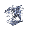 6rqqC 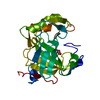 6rquC 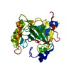 6rqwC  5dvxS S: Starting model for refinement C: citing same article ( |
|---|---|
| Similar structure data |


-Links
 PDBj
PDBj


- Assembly
| Deposited unit | 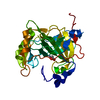
| ||||||||
|---|---|---|---|---|---|---|---|---|---|
| 1 |
| ||||||||
| Unit cell |
|
-Components
| #1: Protein | Mass: 28092.580 Da / Num. of mol.: 1 / Mutation: C174S L183S A213K A258K F259Y M350S Source method: isolated from a genetically manipulated source Source: (gene. exp.) Homo sapiens (human) / Gene: CA9, G250, MN / Production host:  |
|---|---|
| #2: Chemical | ChemComp-ZN /   Mass: 65.409 Da / Num. of mol.: 1 / Source method: obtained synthetically / Formula: Zn Mass: 65.409 Da / Num. of mol.: 1 / Source method: obtained synthetically / Formula: Zn |
| #3: Chemical | ChemComp-ACT /   Mass: 59.044 Da / Num. of mol.: 1 / Source method: obtained synthetically / Formula: C2H3O2 Mass: 59.044 Da / Num. of mol.: 1 / Source method: obtained synthetically / Formula: C2H3O2 |
| #4: Water | ChemComp-HOH /  Mass: 18.015 Da / Num. of mol.: 247 / Source method: isolated from a natural source / Formula: H2O Mass: 18.015 Da / Num. of mol.: 247 / Source method: isolated from a natural source / Formula: H2O |
| Has protein modification | Y |
-Experimental details
-Experiment
| Experiment | Method: X-RAY DIFFRACTION / Number of used crystals: 1 |
|---|
- Sample preparation
| Crystal | Density Matthews: 2.17 Å3/Da / Density % sol: 43.44 % |
|---|---|
| Crystal grow | Temperature: 298 K / Method: vapor diffusion, sitting drop / pH: 8.5 Details: 30% (w/v) PEG 4000, 0.2 M sodium acetate, 0.1 M Tris-HCl pH 8.5 Temp details: ambient |
-Data collection
| Diffraction | Mean temperature: 100 K / Serial crystal experiment: N |
|---|---|
| Diffraction source | Source: SYNCHROTRON / Site: ESRF  / Beamline: BM30A / Wavelength: 0.979 Å / Beamline: BM30A / Wavelength: 0.979 Å |
| Detector | Type: ADSC QUANTUM 315r / Detector: CCD / Date: May 15, 2017 |
| Radiation | Protocol: SINGLE WAVELENGTH / Monochromatic (M) / Laue (L): M / Scattering type: x-ray |
| Radiation wavelength | Wavelength: 0.979 Å / Relative weight: 1 |
| Reflection | Resolution: 1.77→50 Å / Num. obs: 22187 / % possible obs: 95.1 % / Redundancy: 2.8 % / Rmerge(I) obs: 0.063 / Net I/σ(I): 10.5 |
| Reflection shell | Resolution: 1.77→1.88 Å / Redundancy: 2.6 % / Rmerge(I) obs: 0.486 / Mean I/σ(I) obs: 2.2 / Num. unique obs: 3463 / % possible all: 92.9 |
- Processing
| Software |
| |||||||||||||||||||||||||||||||||||||||||||||||||||||||||||||||
|---|---|---|---|---|---|---|---|---|---|---|---|---|---|---|---|---|---|---|---|---|---|---|---|---|---|---|---|---|---|---|---|---|---|---|---|---|---|---|---|---|---|---|---|---|---|---|---|---|---|---|---|---|---|---|---|---|---|---|---|---|---|---|---|---|
| Refinement | Method to determine structure: MOLECULAR REPLACEMENT Starting model: 5dvx Resolution: 1.777→34.171 Å / SU ML: 0.23 / Cross valid method: THROUGHOUT / σ(F): 1.37 / Phase error: 22.78
| |||||||||||||||||||||||||||||||||||||||||||||||||||||||||||||||
| Solvent computation | Shrinkage radii: 0.9 Å / VDW probe radii: 1.11 Å | |||||||||||||||||||||||||||||||||||||||||||||||||||||||||||||||
| Displacement parameters | Biso max: 91.72 Å2 / Biso mean: 27.2776 Å2 / Biso min: 11.19 Å2 | |||||||||||||||||||||||||||||||||||||||||||||||||||||||||||||||
| Refinement step | Cycle: final / Resolution: 1.777→34.171 Å
| |||||||||||||||||||||||||||||||||||||||||||||||||||||||||||||||
| Refine LS restraints |
| |||||||||||||||||||||||||||||||||||||||||||||||||||||||||||||||
| LS refinement shell | Refine-ID: X-RAY DIFFRACTION / Rfactor Rfree error: 0 / Total num. of bins used: 8
|
