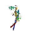
CHAD domain superfamily / CHAD / CHAD domain / CHAD domain / CHAD domain profile. / metal ion binding / CARBONATE ION / COPPER (II) ION / CHAD domain protein
Function and homology information
Biological species
Streptomyces chartreusis (bacteria)
Method
X-RAY DIFFRACTION / SYNCHROTRON / SAD / Resolution: 2.037 Å
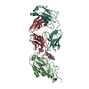
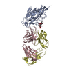
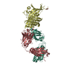
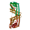
Journal: FEBS Lett / Year: 2019 Title: Structural and biochemical analysis of a phosin from Streptomyces chartreusis reveals a combined polyphosphate- and metal-binding fold. Authors: Sebastiaan Werten / Nils Hinnerk Rustmeier / Maximilian Gemmer / Marie-Joëlle Virolle / Winfried Hinrichs / Abstract: X-ray crystallographic analysis of a phosin (PptA) from Steptomyces chartreusis reveals a metal-associated, lozenge-shaped fold featuring a 5-10 Å wide, positively charged tunnel that traverses the ...X-ray crystallographic analysis of a phosin (PptA) from Steptomyces chartreusis reveals a metal-associated, lozenge-shaped fold featuring a 5-10 Å wide, positively charged tunnel that traverses the protein core. Two distinct metal-binding sites were identified in which the predominant metal ion was Cu . In solution, PptA forms stable homodimers that bind with nanomolar affinity to polyphosphate, a stress-related biopolymer acting as a phosphate and energy reserve in conditions of nutrient depletion. A single protein dimer interacts with 14-15 consecutive phosphate moieties within the polymer. Our observations suggest that PptA plays a role in polyphosphate metabolism, mobilisation or sensing, possibly by acting in concert with polyphosphate kinase (Ppk). Like Ppk, phosins may influence antibiotic synthesis by streptomycetes.
Method to determine structure: SAD / Resolution: 2.037→46.196 Å / Cor.coef. Fo:Fc: 0.962 / Cor.coef. Fo:Fc free: 0.946 / SU B: 5.617 / SU ML: 0.142 / Cross valid method: FREE R-VALUE / ESU R: 0.161 / ESU R Free: 0.155 Details: Hydrogens have been added in their riding positions
Rfactor
Num. reflection
% reflection
Selection details
Rfree
0.237
1228
5 %
Random selection
Rwork
0.191
-
-
-
all
0.193
-
-
-
obs
0.193
24546
98.944 %
-
Solvent computation
Ion probe radii: 0.8 Å / Shrinkage radii: 0.8 Å / VDW probe radii: 1.2 Å
Displacement parameters
Biso mean: 43.16 Å2
Baniso -1
Baniso -2
Baniso -3
1-
5.127 Å2
0 Å2
-0 Å2
2-
-
-2.613 Å2
0 Å2
3-
-
-
-2.515 Å2
Refinement step
Cycle: LAST / Resolution: 2.037→46.196 Å
Protein
Nucleic acid
Ligand
Solvent
Total
Num. atoms
2155
0
28
134
2317
Refine LS restraints
Refine-ID
Type
Dev ideal
Dev ideal target
Number
X-RAY DIFFRACTION
r_bond_refined_d
0.014
0.019
2213
X-RAY DIFFRACTION
r_bond_other_d
0.001
0.02
2112
X-RAY DIFFRACTION
r_angle_refined_deg
1.532
1.962
3008
X-RAY DIFFRACTION
r_angle_other_deg
0.816
3
4827
X-RAY DIFFRACTION
r_dihedral_angle_1_deg
5.552
5
278
X-RAY DIFFRACTION
r_dihedral_angle_2_deg
31.157
20.947
95
X-RAY DIFFRACTION
r_dihedral_angle_3_deg
13.86
15
344
X-RAY DIFFRACTION
r_dihedral_angle_4_deg
17.948
15
30
X-RAY DIFFRACTION
r_chiral_restr
0.08
0.2
354
X-RAY DIFFRACTION
r_gen_planes_refined
0.007
0.021
2454
X-RAY DIFFRACTION
r_gen_planes_other
0.001
0.02
476
X-RAY DIFFRACTION
r_nbd_refined
0.229
0.2
532
X-RAY DIFFRACTION
r_symmetry_nbd_other
0.159
0.2
1975
X-RAY DIFFRACTION
r_nbtor_refined
0.18
0.2
1095
X-RAY DIFFRACTION
r_symmetry_nbtor_other
0.083
0.2
1109
X-RAY DIFFRACTION
r_xyhbond_nbd_refined
0.175
0.2
103
X-RAY DIFFRACTION
r_symmetry_xyhbond_nbd_other
0.055
0.2
2
X-RAY DIFFRACTION
r_metal_ion_refined
0.184
0.2
2
X-RAY DIFFRACTION
r_symmetry_nbd_refined
0.076
0.2
9
X-RAY DIFFRACTION
r_nbd_other
0.136
0.2
37
X-RAY DIFFRACTION
r_symmetry_xyhbond_nbd_refined
0.079
0.2
4
X-RAY DIFFRACTION
r_mcbond_it
3.243
4.063
1127
X-RAY DIFFRACTION
r_mcbond_other
3.239
4.061
1126
X-RAY DIFFRACTION
r_mcangle_it
4.821
6.048
1400
X-RAY DIFFRACTION
r_mcangle_other
4.82
6.051
1401
X-RAY DIFFRACTION
r_scbond_it
4.205
4.623
1086
X-RAY DIFFRACTION
r_scbond_other
4.202
4.604
1068
X-RAY DIFFRACTION
r_scangle_it
6.456
6.756
1608
X-RAY DIFFRACTION
r_scangle_other
6.471
6.726
1582
X-RAY DIFFRACTION
r_lrange_it
8.252
49.385
2546
X-RAY DIFFRACTION
r_lrange_other
8.249
49.267
2521
LS refinement shell
Resolution (Å)
Rfactor Rfree
Num. reflection Rfree
Rfactor Rwork
Num. reflection Rwork
Refine-ID
% reflection obs (%)
2.037-2.09
0.412
82
0.368
1547
X-RAY DIFFRACTION
90.2993
2.09-2.147
0.356
86
0.329
1646
X-RAY DIFFRACTION
97.6875
2.147-2.209
0.264
85
0.273
1603
X-RAY DIFFRACTION
99.8226
2.209-2.277
0.312
83
0.244
1584
X-RAY DIFFRACTION
99.7606
2.277-2.351
0.279
81
0.221
1545
X-RAY DIFFRACTION
99.6324
2.351-2.434
0.239
78
0.192
1475
X-RAY DIFFRACTION
99.7431
2.434-2.525
0.22
75
0.183
1418
X-RAY DIFFRACTION
99.7328
2.525-2.628
0.266
72
0.182
1380
X-RAY DIFFRACTION
99.7938
2.628-2.745
0.211
70
0.185
1334
X-RAY DIFFRACTION
99.7868
2.745-2.878
0.281
67
0.188
1263
X-RAY DIFFRACTION
99.7749
2.878-3.033
0.312
64
0.203
1215
X-RAY DIFFRACTION
100
3.033-3.216
0.243
60
0.19
1148
X-RAY DIFFRACTION
99.8347
3.216-3.437
0.278
57
0.188
1083
X-RAY DIFFRACTION
99.6503
3.437-3.711
0.198
54
0.165
1028
X-RAY DIFFRACTION
100
3.711-4.062
0.237
49
0.156
925
X-RAY DIFFRACTION
99.8974
4.062-4.537
0.191
46
0.151
865
X-RAY DIFFRACTION
100
4.537-5.231
0.206
40
0.164
761
X-RAY DIFFRACTION
99.5031
5.231-6.386
0.215
34
0.202
658
X-RAY DIFFRACTION
100
6.386-8.946
0.176
28
0.142
519
X-RAY DIFFRACTION
99.8175
8.946-46.196
0.153
17
0.185
321
X-RAY DIFFRACTION
99.705
+
About Yorodumi
-
News
-
Feb 9, 2022. New format data for meta-information of EMDB entries
New format data for meta-information of EMDB entries
Version 3 of the EMDB header file is now the official format.
The previous official version 1.9 will be removed from the archive.
In the structure databanks used in Yorodumi, some data are registered as the other names, "COVID-19 virus" and "2019-nCoV". Here are the details of the virus and the list of structure data.
Jan 31, 2019. EMDB accession codes are about to change! (news from PDBe EMDB page)
EMDB accession codes are about to change! (news from PDBe EMDB page)
The allocation of 4 digits for EMDB accession codes will soon come to an end. Whilst these codes will remain in use, new EMDB accession codes will include an additional digit and will expand incrementally as the available range of codes is exhausted. The current 4-digit format prefixed with “EMD-” (i.e. EMD-XXXX) will advance to a 5-digit format (i.e. EMD-XXXXX), and so on. It is currently estimated that the 4-digit codes will be depleted around Spring 2019, at which point the 5-digit format will come into force.
The EM Navigator/Yorodumi systems omit the EMD- prefix.
Related info.:Q: What is EMD? / ID/Accession-code notation in Yorodumi/EM Navigator
Yorodumi is a browser for structure data from EMDB, PDB, SASBDB, etc.
This page is also the successor to EM Navigator detail page, and also detail information page/front-end page for Omokage search.
The word "yorodu" (or yorozu) is an old Japanese word meaning "ten thousand". "mi" (miru) is to see.
Related info.:EMDB / PDB / SASBDB / Comparison of 3 databanks / Yorodumi Search / Aug 31, 2016. New EM Navigator & Yorodumi / Yorodumi Papers / Jmol/JSmol / Function and homology information / Changes in new EM Navigator and Yorodumi
 Movie
Movie Controller


 Open data
 Basic information
 Components
 Keywords
 Function and homology information
 Streptomyces chartreusis (bacteria)
 X-RAY DIFFRACTION /
  Authors
 Citation


 Structure visualization
 Downloads & links
 Other downloads


 PDBj
PDBj


 Assembly
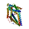


 Mass: 63.546 Da / Num. of mol.: 2 / Source method: obtained synthetically / Formula: Cu / Feature type: SUBJECT OF INVESTIGATION
 Mass: 35.453 Da / Num. of mol.: 2 / Source method: obtained synthetically / Formula: Cl
 Mass: 96.063 Da / Num. of mol.: 4 / Source method: obtained synthetically / Formula: SO4 / Feature type: SUBJECT OF INVESTIGATION
 Mass: 60.009 Da / Num. of mol.: 1 / Source method: obtained synthetically / Formula: CO3
 Sample preparation
 Processing