- SASDF76: Polyphosphate-targeting protein A (PptA) -
+
Open data
ID or keywords:
Loading...
-
Basic information
Entry
Database: SASBDB / ID: SASDF76
Sample
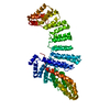
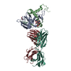
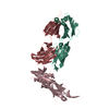
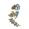
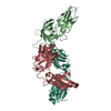
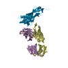
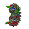
Polyphosphate-targeting protein A
Polyphosphate-targeting protein A (protein), PptA, Streptomyces chartreusis
Function / homology
CHAD domain superfamily / CHAD / CHAD domain / CHAD domain / CHAD domain profile. / metal ion binding / CHAD domain protein
Function and homology information
Biological species
Streptomyces chartreusis (bacteria)
Citation
Journal: FEBS Lett / Year: 2019 Title: Structural and biochemical analysis of a phosin from Streptomyces chartreusis reveals a combined polyphosphate- and metal-binding fold. Authors: Sebastiaan Werten / Nils Hinnerk Rustmeier / Maximilian Gemmer / Marie-Joëlle Virolle / Winfried Hinrichs / Abstract: X-ray crystallographic analysis of a phosin (PptA) from Steptomyces chartreusis reveals a metal-associated, lozenge-shaped fold featuring a 5-10 Å wide, positively charged tunnel that traverses the ...X-ray crystallographic analysis of a phosin (PptA) from Steptomyces chartreusis reveals a metal-associated, lozenge-shaped fold featuring a 5-10 Å wide, positively charged tunnel that traverses the protein core. Two distinct metal-binding sites were identified in which the predominant metal ion was Cu . In solution, PptA forms stable homodimers that bind with nanomolar affinity to polyphosphate, a stress-related biopolymer acting as a phosphate and energy reserve in conditions of nutrient depletion. A single protein dimer interacts with 14-15 consecutive phosphate moieties within the polymer. Our observations suggest that PptA plays a role in polyphosphate metabolism, mobilisation or sensing, possibly by acting in concert with polyphosphate kinase (Ppk). Like Ppk, phosins may influence antibiotic synthesis by streptomycetes.
Contact author
Sebastiaan Werten (Medical University of Innsbruck, Innsbruck, Austria)
Instrument name: PETRA III EMBL P12 / City: Hamburg / 国: Germany / Type of source: X-ray synchrotron / Wavelength: 0.124 Å / Dist. spec. to detc.: 3.1 mm
Detector
Name: Pilatus 2M
Scan
Title: Polyphosphate-targeting protein A / Measurement date: Nov 24, 2016 / Cell temperature: 10 °C / Exposure time: 0.05 sec. / Number of frames: 20 / Unit: 1/nm /
Min
Max
Q
0.0334
3.4982
Distance distribution function P(R)
Sofotware P(R): GNOM 5.0 / Number of points: 1056 /
Min
Max
Q
0.0792223
2.27864
P(R) point
1
1056
R
0
11.84
Result
Type of curve: merged
Experimental
Standard
Porod
MW
78.5 kDa
58.8 kDa
77.3 kDa
Volume
-
-
123.74 nm3
P(R)
Guinier
Guinier error
Forward scattering, I0
4523
4530.06
11.91
Radius of gyration, Rg
3.536 nm
3.51 nm
0.28
Min
Max
D
-
11.84
Guinier point
23
162
+
About Yorodumi
-
News
-
Feb 9, 2022. New format data for meta-information of EMDB entries
New format data for meta-information of EMDB entries
Version 3 of the EMDB header file is now the official format.
The previous official version 1.9 will be removed from the archive.
In the structure databanks used in Yorodumi, some data are registered as the other names, "COVID-19 virus" and "2019-nCoV". Here are the details of the virus and the list of structure data.
Jan 31, 2019. EMDB accession codes are about to change! (news from PDBe EMDB page)
EMDB accession codes are about to change! (news from PDBe EMDB page)
The allocation of 4 digits for EMDB accession codes will soon come to an end. Whilst these codes will remain in use, new EMDB accession codes will include an additional digit and will expand incrementally as the available range of codes is exhausted. The current 4-digit format prefixed with “EMD-” (i.e. EMD-XXXX) will advance to a 5-digit format (i.e. EMD-XXXXX), and so on. It is currently estimated that the 4-digit codes will be depleted around Spring 2019, at which point the 5-digit format will come into force.
The EM Navigator/Yorodumi systems omit the EMD- prefix.
Related info.:Q: What is EMD? / ID/Accession-code notation in Yorodumi/EM Navigator
Yorodumi is a browser for structure data from EMDB, PDB, SASBDB, etc.
This page is also the successor to EM Navigator detail page, and also detail information page/front-end page for Omokage search.
The word "yorodu" (or yorozu) is an old Japanese word meaning "ten thousand". "mi" (miru) is to see.
Related info.:EMDB / PDB / SASBDB / Comparison of 3 databanks / Yorodumi Search / Aug 31, 2016. New EM Navigator & Yorodumi / Yorodumi Papers / Jmol/JSmol / Function and homology information / Changes in new EM Navigator and Yorodumi
 Movie
Movie Controller

 Open data
 Basic information
 Sample
 Function and homology information
 Streptomyces chartreusis (bacteria)
 Citation


 Contact author
 Structure visualization
 Downloads & links
 SASDF76


 Search similar-shape structures of this assembly by Omokage search (details)


