 Movie
Movie Controller

[English] 日本語
 Yorodumi
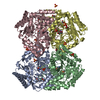
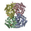
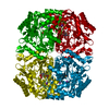
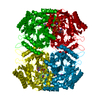
- PDB-6rl5: The first crystal structure of the DABA aminotransferase EctB in ... -
+ Open data

- Basic information
| Entry | Database: PDB / ID: 6rl5 | ||||||
|---|---|---|---|---|---|---|---|
| Title | The first crystal structure of the DABA aminotransferase EctB in the ectoine biosynthesis pathway of the model organism Chromohalobacter salexigens DSM 3034 | ||||||
 Components Components | Diaminobutyrate--2-oxoglutarate transaminase | ||||||
 Keywords Keywords | TRANSFERASE / Ectoine / DABA aminotransferase / osmoadaptation | ||||||
| Function / homology |  Function and homology information Function and homology informationL-2,4-diaminobutyrate:pyruvate transaminase activity / diaminobutyrate-2-oxoglutarate transaminase / L-2,4-diaminobutyrate:2-oxoglutarate transaminase activity / ectoine biosynthetic process / pyridoxal phosphate binding Similarity search - Function | ||||||
| Biological species |  Chromohalobacter salexigens (bacteria) Chromohalobacter salexigens (bacteria) | ||||||
| Method |  X-RAY DIFFRACTION / SYNCHROTRON / MOLECULAR REPLACEMENT / Resolution: 2.45 Å X-RAY DIFFRACTION / SYNCHROTRON / MOLECULAR REPLACEMENT / Resolution: 2.45 Å | ||||||
 Authors Authors | Hillier, H.T. / Altermark, B. / Leiros, I. | ||||||
 Citation Citation | Journal: Febs J. / Year: 2020 Title: The crystal structure of the tetrameric DABA-aminotransferase EctB, a rate-limiting enzyme in the ectoine biosynthesis pathway. Authors: Hillier, H.T. / Altermark, B. / Leiros, I. | ||||||
| History |
|
- Structure visualization
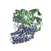
| Structure viewer | Molecule: MolmilJmol/JSmol |
|---|
- Downloads & links
-Download
| PDBx/mmCIF format | 6rl5.cif.gz | 628.5 KB | Display | PDBx/mmCIF format |
|---|---|---|---|---|
| PDB format | pdb6rl5.ent.gz | 520.2 KB | Display | PDB format |
| PDBx/mmJSON format | 6rl5.json.gz | Tree view | PDBx/mmJSON format | |
| Others |  Other downloads Other downloads |
-Validation report
| Arichive directory | https://data.pdbj.org/pub/pdb/validation_reports/rl/6rl5ftp://data.pdbj.org/pub/pdb/validation_reports/rl/6rl5 | HTTPS FTP |
|---|
-Related structure data
| Related structure data |  1sf2S S: Starting model for refinement |
|---|---|
| Similar structure data |
-Links
 PDBj
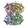
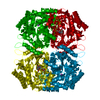
PDBj- Assembly
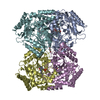
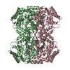
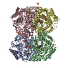
| Deposited unit | 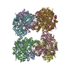
| ||||||||
|---|---|---|---|---|---|---|---|---|---|
| 1 | 
| ||||||||
| 2 | 
| ||||||||
| Unit cell |
|
-Components
| #1: Protein | Mass: 47086.125 Da / Num. of mol.: 8 Source method: isolated from a genetically manipulated source Details: Synthetic construct containing a 6xHIS C-terminal purification tag Source: (gene. exp.) Chromohalobacter salexigens (strain DSM 3043 / ATCC BAA-138 / NCIMB 13768) (bacteria)Strain: DSM 3043 / ATCC BAA-138 / NCIMB 13768 / Gene: ectB, Csal_1877 / Production host: References: UniProt: Q9ZEU7, diaminobutyrate-2-oxoglutarate transaminase #2: Chemical | ChemComp-PLP /   Mass: 247.142 Da / Num. of mol.: 8 / Source method: obtained synthetically / Formula: C8H10NO6P / Feature type: SUBJECT OF INVESTIGATION Mass: 247.142 Da / Num. of mol.: 8 / Source method: obtained synthetically / Formula: C8H10NO6P / Feature type: SUBJECT OF INVESTIGATION#3: Water | ChemComp-HOH / |  Mass: 18.015 Da / Num. of mol.: 310 / Source method: isolated from a natural source / Formula: H2O Mass: 18.015 Da / Num. of mol.: 310 / Source method: isolated from a natural source / Formula: H2O |
|---|
-Experimental details
-Experiment
| Experiment | Method: X-RAY DIFFRACTION / Number of used crystals: 1 |
|---|
- Sample preparation
| Crystal | Density Matthews: 2.55 Å3/Da / Density % sol: 52 % |
|---|---|
| Crystal grow | Temperature: 293 K / Method: vapor diffusion, hanging drop / pH: 6.5 Details: 0.2M sodium malonate, 0.1M BisTris propane pH 6.5, 30% (w/v) PEG 3350 |
-Data collection
| Diffraction | Mean temperature: 100 K / Serial crystal experiment: N |
|---|---|
| Diffraction source | Source: SYNCHROTRON / Site: ESRF  / Beamline: ID23-1 / Wavelength: 0.99187 Å / Beamline: ID23-1 / Wavelength: 0.99187 Å |
| Detector | Type: DECTRIS PILATUS3 S 6M / Detector: PIXEL / Date: Aug 26, 2018 / Details: toroidal mirror |
| Radiation | Monochromator: Si (111) silicon crystal / Protocol: SINGLE WAVELENGTH / Monochromatic (M) / Laue (L): M / Scattering type: x-ray |
| Radiation wavelength | Wavelength: 0.99187 Å / Relative weight: 1 |
| Reflection | Resolution: 2.45→49.073 Å / Num. obs: 136967 / % possible obs: 98.9 % / Observed criterion σ(F): 0 / Observed criterion σ(I): 0 / Redundancy: 3.4 % / Rpim(I) all: 0.047 / Net I/σ(I): 10.6 |
| Reflection shell | Resolution: 2.45→2.49 Å / Redundancy: 3.4 % / Mean I/σ(I) obs: 0.7 / Num. unique obs: 6796 / Rpim(I) all: 1.07 / % possible all: 99.6 |
- Processing
| Software |
| ||||||||||||||||||||||||||||||||||||||||||||||||||||||||||||||||||||||||||||||||||||||||||||||||||||||||||||||||||||||||||||||||||||||||||||
|---|---|---|---|---|---|---|---|---|---|---|---|---|---|---|---|---|---|---|---|---|---|---|---|---|---|---|---|---|---|---|---|---|---|---|---|---|---|---|---|---|---|---|---|---|---|---|---|---|---|---|---|---|---|---|---|---|---|---|---|---|---|---|---|---|---|---|---|---|---|---|---|---|---|---|---|---|---|---|---|---|---|---|---|---|---|---|---|---|---|---|---|---|---|---|---|---|---|---|---|---|---|---|---|---|---|---|---|---|---|---|---|---|---|---|---|---|---|---|---|---|---|---|---|---|---|---|---|---|---|---|---|---|---|---|---|---|---|---|---|---|---|
| Refinement | Method to determine structure: MOLECULAR REPLACEMENT Starting model: 1sf2 Resolution: 2.45→49.073 Å / SU ML: 0.4 / Cross valid method: FREE R-VALUE / σ(F): 1.33 / Phase error: 29.73
| ||||||||||||||||||||||||||||||||||||||||||||||||||||||||||||||||||||||||||||||||||||||||||||||||||||||||||||||||||||||||||||||||||||||||||||
| Solvent computation | Shrinkage radii: 0.9 Å / VDW probe radii: 1.11 Å | ||||||||||||||||||||||||||||||||||||||||||||||||||||||||||||||||||||||||||||||||||||||||||||||||||||||||||||||||||||||||||||||||||||||||||||
| Refinement step | Cycle: LAST / Resolution: 2.45→49.073 Å
| ||||||||||||||||||||||||||||||||||||||||||||||||||||||||||||||||||||||||||||||||||||||||||||||||||||||||||||||||||||||||||||||||||||||||||||
| Refine LS restraints |
| ||||||||||||||||||||||||||||||||||||||||||||||||||||||||||||||||||||||||||||||||||||||||||||||||||||||||||||||||||||||||||||||||||||||||||||
| LS refinement shell |
|
