Movie
Movie Controller


+ Open data

- Basic information
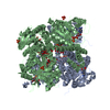
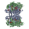
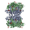
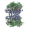
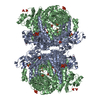
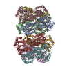
| Entry | Database: PDB / ID: 6rkd | ||||||
|---|---|---|---|---|---|---|---|
| Title | Molybdenum storage protein under turnover conditions | ||||||
 Components Components | (Molybdenum storage protein subunit ...) x 2 | ||||||
 Keywords Keywords | METAL BINDING PROTEIN / molybdenum storage protein / ATPase | ||||||
| Function / homology |  Function and homology information Function and homology information | ||||||
| Biological species |  Azotobacter vinelandii (bacteria) Azotobacter vinelandii (bacteria) | ||||||
| Method | ELECTRON MICROSCOPY / single particle reconstruction / cryo EM / Resolution: 3.2 Å | ||||||
 Authors Authors | Bruenle, S. / Mills, D.J. / Vonck, J. / Ermler, U. | ||||||
 Citation Citation | Journal: Proc Natl Acad Sci U S A / Year: 2019 Title: Molybdate pumping into the molybdenum storage protein via an ATP-powered piercing mechanism. Authors: Steffen Brünle / Martin L Eisinger / Juliane Poppe / Deryck J Mills / Julian D Langer / Janet Vonck / Ulrich Ermler /  Abstract: The molybdenum storage protein (MoSto) deposits large amounts of molybdenum as polyoxomolybdate clusters in a heterohexameric (αβ) cage-like protein complex under ATP consumption. Here, we suggest ...The molybdenum storage protein (MoSto) deposits large amounts of molybdenum as polyoxomolybdate clusters in a heterohexameric (αβ) cage-like protein complex under ATP consumption. Here, we suggest a unique mechanism for the ATP-powered molybdate pumping process based on X-ray crystallography, cryoelectron microscopy, hydrogen-deuterium exchange mass spectrometry, and mutational studies of MoSto from . First, we show that molybdate, ATP, and Mg consecutively bind into the open ATP-binding groove of the β-subunit, which thereafter becomes tightly locked by fixing the previously disordered N-terminal arm of the α-subunit over the β-ATP. Next, we propose a nucleophilic attack of molybdate onto the γ-phosphate of β-ATP, analogous to the similar reaction of the structurally related UMP kinase. The formed instable phosphoric-molybdic anhydride becomes immediately hydrolyzed and, according to the current data, the released and accelerated molybdate is pressed through the cage wall, presumably by turning aside the Metβ149 side chain. A structural comparison between MoSto and UMP kinase provides valuable insight into how an enzyme is converted into a molecular machine during evolution. The postulated direct conversion of chemical energy into kinetic energy via an activating molybdate kinase and an exothermic pyrophosphatase reaction to overcome a proteinous barrier represents a novelty in ATP-fueled biochemistry, because normally, ATP hydrolysis initiates large-scale conformational changes to drive a distant process. | ||||||
| History |
|
- Structure visualization
| Movie |
Movie viewer |
|---|---|
| Structure viewer | Molecule: MolmilJmol/JSmol |
- Downloads & links
-Download
| PDBx/mmCIF format | 6rkd.cif.gz | 622.8 KB | Display | PDBx/mmCIF format |
|---|---|---|---|---|
| PDB format | pdb6rkd.ent.gz | 516.2 KB | Display | PDB format |
| PDBx/mmJSON format | 6rkd.json.gz | Tree view | PDBx/mmJSON format | |
| Others |  Other downloads Other downloads |
-Validation report
| Arichive directory | https://data.pdbj.org/pub/pdb/validation_reports/rk/6rkdftp://data.pdbj.org/pub/pdb/validation_reports/rk/6rkd | HTTPS FTP |
|---|
-Related structure data
| Related structure data |  4907MC  6risC 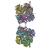 6rj4C 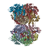 6rkeC M: map data used to model this data C: citing same article ( |
|---|---|
| Similar structure data |


-Links
 PDBj
PDBj- Assembly
| Deposited unit | 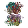
|
|---|---|
| 1 |
|
-Components
-Molybdenum storage protein subunit ... , 2 types, 12 molecules ACEGIKBDFHJL
| #1: Protein | Mass: 29376.773 Da / Num. of mol.: 6 Source method: isolated from a genetically manipulated source Source: (gene. exp.) Azotobacter vinelandii (strain DJ / ATCC BAA-1303) (bacteria)Gene: mosA, Avin_43200 / Production host: #2: Protein | Mass: 28378.775 Da / Num. of mol.: 6 Source method: isolated from a genetically manipulated source Source: (gene. exp.) Azotobacter vinelandii (strain DJ / ATCC BAA-1303) (bacteria)Gene: mosB, Avin_43210 / Production host: |
|---|
-Non-polymers , 6 types, 90 molecules 

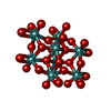
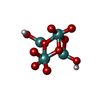


| #3: Chemical | ChemComp-ATP /  Mass: 507.181 Da / Num. of mol.: 12 / Source method: obtained synthetically / Formula: C10H16N5O13P3 / Comment: ATP, energy-carrying molecule*YM Mass: 507.181 Da / Num. of mol.: 12 / Source method: obtained synthetically / Formula: C10H16N5O13P3 / Comment: ATP, energy-carrying molecule*YM#4: Chemical | ChemComp-MG /  Mass: 24.305 Da / Num. of mol.: 6 / Source method: obtained synthetically / Formula: Mg Mass: 24.305 Da / Num. of mol.: 6 / Source method: obtained synthetically / Formula: Mg#5: Chemical | ChemComp-8M0 /  Mass: 1215.503 Da / Num. of mol.: 12 / Source method: obtained synthetically / Formula: Mo8O28 Mass: 1215.503 Da / Num. of mol.: 12 / Source method: obtained synthetically / Formula: Mo8O28#6: Chemical | ChemComp-J8E /  Mass: 545.770 Da / Num. of mol.: 12 / Source method: obtained synthetically / Formula: H2Mo4O10 Mass: 545.770 Da / Num. of mol.: 12 / Source method: obtained synthetically / Formula: H2Mo4O10#7: Chemical | ChemComp-MOO /  Mass: 159.938 Da / Num. of mol.: 42 / Source method: obtained synthetically / Formula: MoO4 Mass: 159.938 Da / Num. of mol.: 42 / Source method: obtained synthetically / Formula: MoO4#8: Chemical | ChemComp-OMO /  Mass: 145.954 Da / Num. of mol.: 6 / Source method: obtained synthetically / Formula: H2MoO3 Mass: 145.954 Da / Num. of mol.: 6 / Source method: obtained synthetically / Formula: H2MoO3 |
|---|
-Experimental details
-Experiment
| Experiment | Method: ELECTRON MICROSCOPY |
|---|---|
| EM experiment | Aggregation state: PARTICLE / 3D reconstruction method: single particle reconstruction |
- Sample preparation
| Component | Name: Dimer of A3B3 heterohexamer of molybdenum storage protein Type: COMPLEX / Entity ID: #1-#2 / Source: RECOMBINANT |
|---|---|
| Molecular weight | Value: 0.38 MDa / Experimental value: NO |
| Source (natural) | Organism: Azotobacter vinelandii DJ (bacteria) |
| Source (recombinant) | Organism: |
| Buffer solution | pH: 6.5 Details: 1 mM molybdate and 1 mM mg-ATP were added before vitrification. |
| Buffer component | Conc.: 50 mM / Name: MOPS/NaOH |
| Specimen | Conc.: 4 mg/ml / Embedding applied: NO / Shadowing applied: NO / Staining applied: NO / Vitrification applied: YES |
| Specimen support | Grid material: COPPER / Grid type: C-flat |
| Vitrification | Instrument: FEI VITROBOT MARK IV / Cryogen name: ETHANE / Humidity: 70 % / Chamber temperature: 283 K |
- Electron microscopy imaging
| Microscopy | Model: JEOL 3200FSC |
|---|---|
| Electron gun | Electron source:  FIELD EMISSION GUN / Accelerating voltage: 300 kV / Illumination mode: FLOOD BEAM FIELD EMISSION GUN / Accelerating voltage: 300 kV / Illumination mode: FLOOD BEAM |
| Electron lens | Mode: BRIGHT FIELD / Nominal magnification: 30000 X / Calibrated magnification: 45045 X / Calibrated defocus min: 900 nm / Calibrated defocus max: 2500 nm / Cs: 2.7 mm / Alignment procedure: COMA FREE |
| Specimen holder | Cryogen: NITROGEN / Specimen holder model: JEOL |
| Image recording | Average exposure time: 8 sec. / Electron dose: 54 e/Å2 / Detector mode: COUNTING / Film or detector model: GATAN K2 SUMMIT (4k x 4k) / Num. of real images: 1238 |
| EM imaging optics | Energyfilter name: In-column Omega Filter / Energyfilter slit width: 20 eV |
| Image scans | Movie frames/image: 40 |
- Processing
| EM software |
| ||||||||||||||||||||||||||||||||
|---|---|---|---|---|---|---|---|---|---|---|---|---|---|---|---|---|---|---|---|---|---|---|---|---|---|---|---|---|---|---|---|---|---|
| CTF correction | Type: PHASE FLIPPING ONLY | ||||||||||||||||||||||||||||||||
| Particle selection | Num. of particles selected: 174681 | ||||||||||||||||||||||||||||||||
| Symmetry | Point symmetry: D3 (2x3 fold dihedral) | ||||||||||||||||||||||||||||||||
| 3D reconstruction | Resolution: 3.2 Å / Resolution method: FSC 0.143 CUT-OFF / Num. of particles: 137558 / Symmetry type: POINT | ||||||||||||||||||||||||||||||||
| Atomic model building | Protocol: OTHER / Space: REAL / Details: Phenix_real_space_refine | ||||||||||||||||||||||||||||||||
| Atomic model building | 3D fitting-ID: 1 / Accession code: 4F6T / Initial refinement model-ID: 1 / PDB-ID: 4F6T / Source name: PDB / Type: experimental model
|