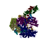
 Movie
Movie Controller

[English] 日本語
 Yorodumi
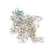
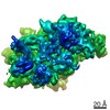
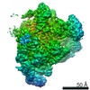
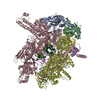
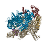
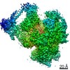
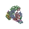
- PDB-6r9b: Cryo-EM structure of bacterial RNAP with a DNA mimic protein Ocr ... -
+ Open data

- Basic information
| Entry | Database: PDB / ID: 6r9b | ||||||
|---|---|---|---|---|---|---|---|
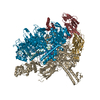
| Title | Cryo-EM structure of bacterial RNAP with a DNA mimic protein Ocr from T7 phage | ||||||
 Components Components |
| ||||||
 Keywords Keywords | TRANSCRIPTION / RNA polymerase / Ocr / transcription inhibition / bacteriophage T7 | ||||||
| Function / homology |  Function and homology information Function and homology informationsymbiont-mediated evasion of host restriction-modification system / RNA polymerase complex / submerged biofilm formation / cellular response to cell envelope stress / regulation of DNA-templated transcription initiation / bacterial-type flagellum assembly / bacterial-type RNA polymerase core enzyme binding / cytosolic DNA-directed RNA polymerase complex / bacterial-type flagellum-dependent cell motility / nitrate assimilation ...symbiont-mediated evasion of host restriction-modification system / RNA polymerase complex / submerged biofilm formation / cellular response to cell envelope stress / regulation of DNA-templated transcription initiation / bacterial-type flagellum assembly / bacterial-type RNA polymerase core enzyme binding / cytosolic DNA-directed RNA polymerase complex / bacterial-type flagellum-dependent cell motility / nitrate assimilation / regulation of DNA-templated transcription elongation / transcription elongation factor complex / DNA-directed RNA polymerase complex / transcription antitermination / DNA-templated transcription initiation / cell motility / ribonucleoside binding / DNA-directed RNA polymerase / DNA-directed RNA polymerase activity / response to heat / protein-containing complex assembly / intracellular iron ion homeostasis / protein dimerization activity / symbiont-mediated suppression of host innate immune response / response to antibiotic / DNA-templated transcription / magnesium ion binding / DNA binding / zinc ion binding / membrane / cytoplasm / cytosol Similarity search - Function | ||||||
| Biological species |    Enterobacteria phage T7 (virus) Enterobacteria phage T7 (virus) | ||||||
| Method | ELECTRON MICROSCOPY / single particle reconstruction / cryo EM / Resolution: 3.8 Å | ||||||
 Authors Authors | Ye, F.Z. / Zhang, X.D. | ||||||
| Funding support |  United Kingdom, 1items United Kingdom, 1items
| ||||||
 Citation Citation | Journal: Elife / Year: 2020 Title: Structural basis of transcription inhibition by the DNA mimic protein Ocr of bacteriophage T7. Authors: Fuzhou Ye / Ioly Kotta-Loizou / Milija Jovanovic / Xiaojiao Liu / David Tf Dryden / Martin Buck / Xiaodong Zhang /  Abstract: Bacteriophage T7 infects and evades the host restriction/modification system. The Ocr protein of T7 was shown to exist as a dimer mimicking DNA and to bind to host restriction enzymes, thus ...Bacteriophage T7 infects and evades the host restriction/modification system. The Ocr protein of T7 was shown to exist as a dimer mimicking DNA and to bind to host restriction enzymes, thus preventing the degradation of the viral genome by the host. Here we report that Ocr can also inhibit host transcription by directly binding to bacterial RNA polymerase (RNAP) and competing with the recruitment of RNAP by sigma factors. Using cryo electron microscopy, we determined the structures of Ocr bound to RNAP. The structures show that an Ocr dimer binds to RNAP in the cleft, where key regions of sigma bind and where DNA resides during transcription synthesis, thus providing a structural basis for the transcription inhibition. Our results reveal the versatility of Ocr in interfering with host systems and suggest possible strategies that could be exploited in adopting DNA mimicry as a basis for forming novel antibiotics. | ||||||
| History |
|
- Structure visualization
| Movie |
Movie viewer |
|---|---|
| Structure viewer | Molecule: MolmilJmol/JSmol |
- Downloads & links
-Download
| PDBx/mmCIF format | 6r9b.cif.gz | 587.9 KB | Display | PDBx/mmCIF format |
|---|---|---|---|---|
| PDB format | pdb6r9b.ent.gz | 469.6 KB | Display | PDB format |
| PDBx/mmJSON format | 6r9b.json.gz | Tree view | PDBx/mmJSON format | |
| Others |  Other downloads Other downloads |
-Validation report
| Arichive directory | https://data.pdbj.org/pub/pdb/validation_reports/r9/6r9bftp://data.pdbj.org/pub/pdb/validation_reports/r9/6r9b | HTTPS FTP |
|---|
-Related structure data
| Related structure data | 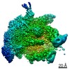 4769MC 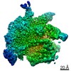 4770C 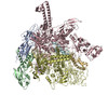 6r9gC M: map data used to model this data C: citing same article ( |
|---|---|
| Similar structure data |

-Links
 PDBj
PDBj


- Assembly
| Deposited unit | 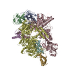
|
|---|---|
| 1 |
|
-Components
| #1: Protein | Mass: 36558.680 Da / Num. of mol.: 2 Source method: isolated from a genetically manipulated source Source: (gene. exp.) Strain: K12 / Gene: rpoA, pez, phs, sez, b3295, JW3257 / Plasmid: pGEM-ABC / Production host: #2: Protein | | Mass: 150820.875 Da / Num. of mol.: 1 Source method: isolated from a genetically manipulated source Source: (gene. exp.) Strain: K12 Gene: rpoB, groN, nitB, rif, ron, stl, stv, tabD, b3987, JW3950 Plasmid: pGEMABC / Production host: #3: Protein | | Mass: 155366.781 Da / Num. of mol.: 1 Source method: isolated from a genetically manipulated source Source: (gene. exp.) Strain: K12 / Gene: rpoC, tabB, b3988, JW3951 / Plasmid: pGEMABC / Production host: #4: Protein | | Mass: 9094.239 Da / Num. of mol.: 1 Source method: isolated from a genetically manipulated source Source: (gene. exp.) References: UniProt: F4NQ47, UniProt: P0A800*PLUS, DNA-directed RNA polymerase #5: Protein | Mass: 13819.015 Da / Num. of mol.: 2 Source method: isolated from a genetically manipulated source Source: (gene. exp.) Enterobacteria phage T7 (virus) / Gene: 0.3 / Plasmid: pOPINF-Ocr / Production host: Has ligand of interest | Y | Has protein modification | Y | |
|---|
-Experimental details
-Experiment
| Experiment | Method: ELECTRON MICROSCOPY |
|---|---|
| EM experiment | Aggregation state: PARTICLE / 3D reconstruction method: single particle reconstruction |
- Sample preparation
| Component |
| |||||||||||||||||||||||||
|---|---|---|---|---|---|---|---|---|---|---|---|---|---|---|---|---|---|---|---|---|---|---|---|---|---|---|
| Molecular weight | Value: 0.410 MDa / Experimental value: NO | |||||||||||||||||||||||||
| Source (natural) |
| |||||||||||||||||||||||||
| Source (recombinant) |
| |||||||||||||||||||||||||
| Buffer solution | pH: 8 | |||||||||||||||||||||||||
| Buffer component |
| |||||||||||||||||||||||||
| Specimen | Conc.: 0.6 mg/ml / Embedding applied: NO / Shadowing applied: NO / Staining applied: NO / Vitrification applied: YES | |||||||||||||||||||||||||
| Specimen support | Grid mesh size: 200 divisions/in. / Grid type: Quantifoil R2/2 | |||||||||||||||||||||||||
| Vitrification | Instrument: FEI VITROBOT MARK IV / Cryogen name: ETHANE / Humidity: 95 % / Chamber temperature: 277 K |
- Electron microscopy imaging
| Experimental equipment |  Model: Titan Krios / Image courtesy: FEI Company |
|---|---|
| Microscopy | Model: FEI TITAN KRIOS |
| Electron gun | Electron source:  FIELD EMISSION GUN / Accelerating voltage: 300 kV / Illumination mode: OTHER FIELD EMISSION GUN / Accelerating voltage: 300 kV / Illumination mode: OTHER |
| Electron lens | Mode: BRIGHT FIELD / Nominal defocus max: -3 nm / Nominal defocus min: -1.2 nm |
| Image recording | Average exposure time: 1 sec. / Electron dose: 49.53 e/Å2 / Detector mode: COUNTING / Film or detector model: GATAN K2 SUMMIT (4k x 4k) / Num. of grids imaged: 1 / Num. of real images: 3543 |
| Image scans | Movie frames/image: 41 |
- Processing
| Software | Name: PHENIX / Version: 1.13_2998: / Classification: refinement | ||||||||||||||||||||||||
|---|---|---|---|---|---|---|---|---|---|---|---|---|---|---|---|---|---|---|---|---|---|---|---|---|---|
| EM software |
| ||||||||||||||||||||||||
| CTF correction | Type: PHASE FLIPPING AND AMPLITUDE CORRECTION | ||||||||||||||||||||||||
| Particle selection | Num. of particles selected: 753783 | ||||||||||||||||||||||||
| Symmetry | Point symmetry: C1 (asymmetric) | ||||||||||||||||||||||||
| 3D reconstruction | Resolution: 3.8 Å / Resolution method: FSC 0.143 CUT-OFF / Num. of particles: 27312 / Num. of class averages: 1 / Symmetry type: POINT | ||||||||||||||||||||||||
| Atomic model building | Protocol: RIGID BODY FIT / Space: REAL | ||||||||||||||||||||||||
| Refine LS restraints |
|
