 Movie
Movie Controller

[English] 日本語
 Yorodumi
- PDB-6r76: Crystal structure of trans-3-Hydroxy-L-proline dehydratase from T... -
+ Open data

- Basic information
| Entry | Database: PDB / ID: 6r76 | ||||||
|---|---|---|---|---|---|---|---|
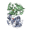
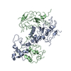
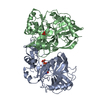
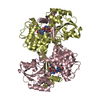
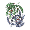
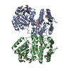
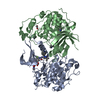
| Title | Crystal structure of trans-3-Hydroxy-L-proline dehydratase from Thermococcus litoralis - open conformation | ||||||
 Components Components | (Proline racemase) x 2 | ||||||
 Keywords Keywords | LYASE / Hydro-lyase / hydroxyproline dehydratase | ||||||
| Function / homology | 4-hydroxyproline epimerase activity / Proline racemase family / Proline racemase / Diaminopimelate Epimerase; Chain A, domain 1 / Diaminopimelate Epimerase; Chain A, domain 1 / Roll / Alpha Beta / Proline racemase Function and homology information Function and homology information | ||||||
| Biological species |   Thermococcus litoralis DSM 5473 (archaea) Thermococcus litoralis DSM 5473 (archaea) | ||||||
| Method |  X-RAY DIFFRACTION / SYNCHROTRON / MOLECULAR REPLACEMENT / Resolution: 2.4 Å X-RAY DIFFRACTION / SYNCHROTRON / MOLECULAR REPLACEMENT / Resolution: 2.4 Å | ||||||
 Authors Authors | Ferraris, D.M. / Miggiano, R. / Rizzi, M. | ||||||
 Citation Citation | Journal: Biochem.Biophys.Res.Commun. / Year: 2019 Title: Structure of Thermococcus litoralis trans-3-hydroxy-l-proline dehydratase in the free and substrate-complexed form. Authors: Ferraris, D.M. / Miggiano, R. / Watanabe, S. / Rizzi, M. | ||||||
| History |
|
- Structure visualization
| Structure viewer | Molecule: MolmilJmol/JSmol |
|---|
- Downloads & links
-Download
| PDBx/mmCIF format | 6r76.cif.gz | 274.7 KB | Display | PDBx/mmCIF format |
|---|---|---|---|---|
| PDB format | pdb6r76.ent.gz | 222.2 KB | Display | PDB format |
| PDBx/mmJSON format | 6r76.json.gz | Tree view | PDBx/mmJSON format | |
| Others |  Other downloads Other downloads |
-Validation report
| Arichive directory | https://data.pdbj.org/pub/pdb/validation_reports/r7/6r76ftp://data.pdbj.org/pub/pdb/validation_reports/r7/6r76 | HTTPS FTP |
|---|
-Related structure data
| Related structure data | 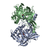 6r77C  1w61S S: Starting model for refinement C: citing same article ( |
|---|---|
| Similar structure data |

-Links
 PDBj
PDBj- Assembly
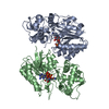
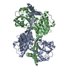
| Deposited unit | 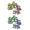
| ||||||||
|---|---|---|---|---|---|---|---|---|---|
| 1 | 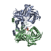
| ||||||||
| 2 | 
| ||||||||
| Unit cell |
|
-Components
| #1: Protein | Mass: 41675.664 Da / Num. of mol.: 2 Source method: isolated from a genetically manipulated source Details: The construct carries an N-term 6xHis-tag and a TEV cleavage site Source: (gene. exp.) Thermococcus litoralis DSM 5473 (archaea)Gene: OCC_00387 / Variant: ATCC 51850 / DSM 5473 / JCM 8560 / NS-C / Production host:  #2: Protein | Mass: 41755.641 Da / Num. of mol.: 2 Source method: isolated from a genetically manipulated source Details: The construct carries an N-term 6xHis-tag and a TEV cleavage site Source: (gene. exp.) Thermococcus litoralis DSM 5473 (archaea)Gene: OCC_00387 / Variant: ATCC 51850 / DSM 5473 / JCM 8560 / NS-C / Production host: #3: Water | ChemComp-HOH / |  Mass: 18.015 Da / Num. of mol.: 16 / Source method: isolated from a natural source / Formula: H2O Mass: 18.015 Da / Num. of mol.: 16 / Source method: isolated from a natural source / Formula: H2O |
|---|
-Experimental details
-Experiment
| Experiment | Method: X-RAY DIFFRACTION / Number of used crystals: 1 |
|---|
- Sample preparation
| Crystal | Density Matthews: 2.16 Å3/Da / Density % sol: 43.09 % |
|---|---|
| Crystal grow | Temperature: 298 K / Method: vapor diffusion, sitting drop / Details: 0.1 M Magnesium formate, 15% PEG3350 |
-Data collection
| Diffraction | Mean temperature: 100 K / Serial crystal experiment: N |
|---|---|
| Diffraction source | Source: SYNCHROTRON / Site: ESRF  / Beamline: ID30B / Wavelength: 0.979163 Å / Beamline: ID30B / Wavelength: 0.979163 Å |
| Detector | Type: DECTRIS PILATUS3 6M / Detector: PIXEL / Date: Feb 26, 2018 |
| Radiation | Protocol: SINGLE WAVELENGTH / Monochromatic (M) / Laue (L): M / Scattering type: x-ray |
| Radiation wavelength | Wavelength: 0.979163 Å / Relative weight: 1 |
| Reflection | Resolution: 2.4→44.8 Å / Num. obs: 53298 / % possible obs: 99.3 % / Redundancy: 1.9 % / Rmerge(I) obs: 0.063 / Net I/σ(I): 7.6 |
| Reflection shell | Resolution: 2.4→2.5 Å / Rmerge(I) obs: 0.41 / Num. unique obs: 5281 |
- Processing
| Software |
| ||||||||||||||||||||||||||||||||||||||||||||||||||||||||||||||||||||||||||||||||||||||||||||||||||||||||||||||||||||||||||||||||||||||||||||
|---|---|---|---|---|---|---|---|---|---|---|---|---|---|---|---|---|---|---|---|---|---|---|---|---|---|---|---|---|---|---|---|---|---|---|---|---|---|---|---|---|---|---|---|---|---|---|---|---|---|---|---|---|---|---|---|---|---|---|---|---|---|---|---|---|---|---|---|---|---|---|---|---|---|---|---|---|---|---|---|---|---|---|---|---|---|---|---|---|---|---|---|---|---|---|---|---|---|---|---|---|---|---|---|---|---|---|---|---|---|---|---|---|---|---|---|---|---|---|---|---|---|---|---|---|---|---|---|---|---|---|---|---|---|---|---|---|---|---|---|---|---|
| Refinement | Method to determine structure: MOLECULAR REPLACEMENT Starting model: 1W61 Resolution: 2.4→44.8 Å / SU ML: 0.35 / Cross valid method: FREE R-VALUE / σ(F): 1.33 / Phase error: 30.92
| ||||||||||||||||||||||||||||||||||||||||||||||||||||||||||||||||||||||||||||||||||||||||||||||||||||||||||||||||||||||||||||||||||||||||||||
| Solvent computation | Shrinkage radii: 0.9 Å / VDW probe radii: 1.11 Å | ||||||||||||||||||||||||||||||||||||||||||||||||||||||||||||||||||||||||||||||||||||||||||||||||||||||||||||||||||||||||||||||||||||||||||||
| Refinement step | Cycle: LAST / Resolution: 2.4→44.8 Å
| ||||||||||||||||||||||||||||||||||||||||||||||||||||||||||||||||||||||||||||||||||||||||||||||||||||||||||||||||||||||||||||||||||||||||||||
| Refine LS restraints |
| ||||||||||||||||||||||||||||||||||||||||||||||||||||||||||||||||||||||||||||||||||||||||||||||||||||||||||||||||||||||||||||||||||||||||||||
| LS refinement shell |
|
