 Movie
Movie Controller


+ Open data

- Basic information
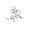
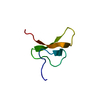
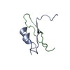
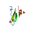
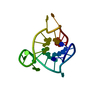
| Entry | Database: PDB / ID: 6r5m | |||||||||
|---|---|---|---|---|---|---|---|---|---|---|
| Title | Crystal structure of toxin MT9 from mamba venom | |||||||||
 Components Components | Dendroaspis polylepis MT9 | |||||||||
 Keywords Keywords | TOXIN / mamba venom / aminergic toxin | |||||||||
| Function / homology |  Function and homology information Function and homology information | |||||||||
| Biological species |  Dendroaspis polylepis (cobra) Dendroaspis polylepis (cobra) | |||||||||
| Method |  X-RAY DIFFRACTION / SYNCHROTRON / MOLECULAR REPLACEMENT / Resolution: 1.9 Å X-RAY DIFFRACTION / SYNCHROTRON / MOLECULAR REPLACEMENT / Resolution: 1.9 Å | |||||||||
 Authors Authors | Stura, E.A. / Tepshi, L. / Ciolek, J. / Triquigneaux, M. / Zoukimian, C. / De Waard, M. / Beroud, R. / Servent, D. / Gilles, N. / Legrand, P. / Ciccone, L. | |||||||||
| Funding support |  France, 1items France, 1items
| |||||||||
 Citation Citation | Journal: Biomed Pharmacother / Year: 2022 Title: MT9, a natural peptide from black mamba venom antagonizes the muscarinic type 2 receptor and reverses the M2R-agonist-induced relaxation in rat and human arteries Authors: Ciolek, J. / Zoukimian, C. / Dhot, J. / Burban, M. / Triquigneaux, M. / Lauzier, B. / Guimbert, C. / Boturyn, D. / Ferron, M. / Ciccone, L. / Tepshi, L. / Stura, E. / Legrand, P. / Robin, P. ...Authors: Ciolek, J. / Zoukimian, C. / Dhot, J. / Burban, M. / Triquigneaux, M. / Lauzier, B. / Guimbert, C. / Boturyn, D. / Ferron, M. / Ciccone, L. / Tepshi, L. / Stura, E. / Legrand, P. / Robin, P. / Mourier, G. / Schaack, B. / Fellah, I. / Blanchet, G. / Gauthier-Erfanian, C. / Beroud, R. / Servent, D. / De Waard, M. / Gilles, N. | |||||||||
| History |
|
- Structure visualization
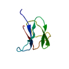
| Structure viewer | Molecule: MolmilJmol/JSmol |
|---|
- Downloads & links
-Download
| PDBx/mmCIF format | 6r5m.cif.gz | 82.5 KB | Display | PDBx/mmCIF format |
|---|---|---|---|---|
| PDB format | pdb6r5m.ent.gz | 66.9 KB | Display | PDB format |
| PDBx/mmJSON format | 6r5m.json.gz | Tree view | PDBx/mmJSON format | |
| Others |  Other downloads Other downloads |
-Validation report
| Arichive directory | https://data.pdbj.org/pub/pdb/validation_reports/r5/6r5mftp://data.pdbj.org/pub/pdb/validation_reports/r5/6r5m | HTTPS FTP |
|---|
-Related structure data
| Similar structure data |
|---|

-Links
 PDBj
PDBj

- Assembly
| Deposited unit | 
| |||||||||
|---|---|---|---|---|---|---|---|---|---|---|
| 1 | 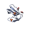
| |||||||||
| 2 | 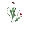
| |||||||||
| 3 | 
| |||||||||
| Unit cell |
| |||||||||
| Components on special symmetry positions |
|
-Components
-Protein , 1 types, 3 molecules ABC
| #1: Protein | Mass: 6374.473 Da / Num. of mol.: 3 / Source method: obtained synthetically / Source: (synth.) Dendroaspis polylepis (cobra) / References: UniProt: A0A4P1LYC9*PLUS |
|---|
-Non-polymers , 5 types, 168 molecules 


| #2: Chemical | ChemComp-SO4 /  Mass: 96.063 Da / Num. of mol.: 9 / Source method: obtained synthetically / Formula: SO4 Mass: 96.063 Da / Num. of mol.: 9 / Source method: obtained synthetically / Formula: SO4#3: Chemical |  Mass: 76.094 Da / Num. of mol.: 2 / Source method: isolated from a natural source / Formula: C3H8O2 Mass: 76.094 Da / Num. of mol.: 2 / Source method: isolated from a natural source / Formula: C3H8O2#4: Chemical | ChemComp-GOL / |  Mass: 92.094 Da / Num. of mol.: 1 / Source method: obtained synthetically / Formula: C3H8O3 Mass: 92.094 Da / Num. of mol.: 1 / Source method: obtained synthetically / Formula: C3H8O3#5: Chemical | ChemComp-ACE / |  Mass: 44.053 Da / Num. of mol.: 1 / Source method: obtained synthetically / Formula: C2H4O Mass: 44.053 Da / Num. of mol.: 1 / Source method: obtained synthetically / Formula: C2H4O#6: Water | ChemComp-HOH / | Mass: 18.015 Da / Num. of mol.: 155 / Source method: isolated from a natural source / Formula: H2O |
|---|
-Experimental details
-Experiment
| Experiment | Method: X-RAY DIFFRACTION / Number of used crystals: 1 |
|---|
- Sample preparation
| Crystal | Density Matthews: 2.65 Å3/Da / Density % sol: 53.61 % |
|---|---|
| Crystal grow | Temperature: 293 K / Method: vapor diffusion, sitting drop / pH: 5.5 Details: 1.05 M A.S 0.06 M sodium citrate, pH 5.5 with 35 mM NaSCN. |
-Data collection
| Diffraction | Mean temperature: 100 K / Serial crystal experiment: N |
|---|---|
| Diffraction source | Source: SYNCHROTRON / Site: SOLEIL / Beamline: PROXIMA 2 / Wavelength: 0.980097 Å |
| Detector | Type: DECTRIS EIGER X 9M / Detector: PIXEL / Date: Sep 20, 2017 / Details: KB Mirrors |
| Radiation | Monochromator: [111]Si Cut monochromator / Protocol: SINGLE WAVELENGTH / Monochromatic (M) / Laue (L): M / Scattering type: x-ray |
| Radiation wavelength | Wavelength: 0.980097 Å / Relative weight: 1 |
| Reflection | Resolution: 1.9→47 Å / Num. obs: 16308 / % possible obs: 99.7 % / Observed criterion σ(F): 0 / Observed criterion σ(I): 0 / Redundancy: 6 % / Biso Wilson estimate: 34.42 Å2 / CC1/2: 0.99 / Rmerge(I) obs: 0.177 / Rpim(I) all: 0.078 / Rrim(I) all: 0.199 / Net I/σ(I): 6.6 |
| Reflection shell | Highest resolution: 1.9 Å |
- Processing
| Software |
| ||||||||||||||||||||||||||||||||||||||||||||||||||||||||||||||||||||||||||||||||||||||||||||||||||||||||||||||||||
|---|---|---|---|---|---|---|---|---|---|---|---|---|---|---|---|---|---|---|---|---|---|---|---|---|---|---|---|---|---|---|---|---|---|---|---|---|---|---|---|---|---|---|---|---|---|---|---|---|---|---|---|---|---|---|---|---|---|---|---|---|---|---|---|---|---|---|---|---|---|---|---|---|---|---|---|---|---|---|---|---|---|---|---|---|---|---|---|---|---|---|---|---|---|---|---|---|---|---|---|---|---|---|---|---|---|---|---|---|---|---|---|---|---|---|---|
| Refinement | Method to determine structure: MOLECULAR REPLACEMENT / Resolution: 1.9→19.45 Å / Cor.coef. Fo:Fc: 0.913 / Cor.coef. Fo:Fc free: 0.909 / SU R Cruickshank DPI: 0.17 / Cross valid method: THROUGHOUT / σ(F): 0 / SU R Blow DPI: 0.192 / SU Rfree Blow DPI: 0.156 / SU Rfree Cruickshank DPI: 0.146
| ||||||||||||||||||||||||||||||||||||||||||||||||||||||||||||||||||||||||||||||||||||||||||||||||||||||||||||||||||
| Displacement parameters | Biso mean: 40.18 Å2
| ||||||||||||||||||||||||||||||||||||||||||||||||||||||||||||||||||||||||||||||||||||||||||||||||||||||||||||||||||
| Refine analyze | Luzzati coordinate error obs: 0.34 Å | ||||||||||||||||||||||||||||||||||||||||||||||||||||||||||||||||||||||||||||||||||||||||||||||||||||||||||||||||||
| Refinement step | Cycle: LAST / Resolution: 1.9→19.45 Å
| ||||||||||||||||||||||||||||||||||||||||||||||||||||||||||||||||||||||||||||||||||||||||||||||||||||||||||||||||||
| Refine LS restraints |
| ||||||||||||||||||||||||||||||||||||||||||||||||||||||||||||||||||||||||||||||||||||||||||||||||||||||||||||||||||
| LS refinement shell | Resolution: 1.9→1.92 Å / Total num. of bins used: 40
| ||||||||||||||||||||||||||||||||||||||||||||||||||||||||||||||||||||||||||||||||||||||||||||||||||||||||||||||||||
| Refinement TLS params. | Method: refined / Refine-ID: X-RAY DIFFRACTION
| ||||||||||||||||||||||||||||||||||||||||||||||||||||||||||||||||||||||||||||||||||||||||||||||||||||||||||||||||||
| Refinement TLS group |
|
