 Movie
Movie Controller

[English] 日本語
 Yorodumi
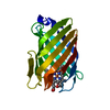
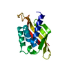
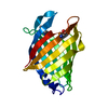
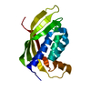
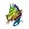
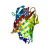
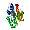
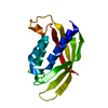
- PDB-6r3y: M.tuberculosis nitrobindin with a cyanide molecule coordinated to... -
+ Open data

- Basic information
| Entry | Database: PDB / ID: 6r3y | ||||||
|---|---|---|---|---|---|---|---|
| Title | M.tuberculosis nitrobindin with a cyanide molecule coordinated to the heme iron atom | ||||||
 Components Components | UPF0678 fatty acid-binding protein-like protein ERS007657_00996 | ||||||
 Keywords Keywords | PROTEIN BINDING / hemeproteins / beta-barrel / peroxynitrite detoxification | ||||||
| Function / homology |  Function and homology information Function and homology informationperoxynitrite isomerase activity / Isomerases; Other isomerases / heme binding / metal ion binding Similarity search - Function | ||||||
| Biological species |   Mycobacterium tuberculosis (bacteria) Mycobacterium tuberculosis (bacteria) | ||||||
| Method |  X-RAY DIFFRACTION / SYNCHROTRON / MOLECULAR REPLACEMENT / Resolution: 1.6 Å X-RAY DIFFRACTION / SYNCHROTRON / MOLECULAR REPLACEMENT / Resolution: 1.6 Å | ||||||
 Authors Authors | De Simone, G. / di Masi, A. / Polticelli, F. / Pesce, A. / Nardini, M. / Bolognesi, M. / Ciaccio, C. / Coletta, M. / Turilli, E.S. / Fasano, M. ...De Simone, G. / di Masi, A. / Polticelli, F. / Pesce, A. / Nardini, M. / Bolognesi, M. / Ciaccio, C. / Coletta, M. / Turilli, E.S. / Fasano, M. / Tognaccini, L. / Smulevich, G. / Abbruzzetti, S. / Viappiani, C. / Bruno, S. / Ascenzi, P. | ||||||
 Citation Citation | Journal: Antioxid.Redox Signal. / Year: 2020 Title: Mycobacterial and Human Nitrobindins: Structure and Function. Authors: De Simone, G. / di Masi, A. / Vita, G.M. / Polticelli, F. / Pesce, A. / Nardini, M. / Bolognesi, M. / Ciaccio, C. / Coletta, M. / Turilli, E.S. / Fasano, M. / Tognaccini, L. / Smulevich, G. ...Authors: De Simone, G. / di Masi, A. / Vita, G.M. / Polticelli, F. / Pesce, A. / Nardini, M. / Bolognesi, M. / Ciaccio, C. / Coletta, M. / Turilli, E.S. / Fasano, M. / Tognaccini, L. / Smulevich, G. / Abbruzzetti, S. / Viappiani, C. / Bruno, S. / Ascenzi, P. | ||||||
| History |
|
- Structure visualization
| Structure viewer | Molecule: MolmilJmol/JSmol |
|---|
- Downloads & links
-Download
| PDBx/mmCIF format | 6r3y.cif.gz | 49.4 KB | Display | PDBx/mmCIF format |
|---|---|---|---|---|
| PDB format | pdb6r3y.ent.gz | 33 KB | Display | PDB format |
| PDBx/mmJSON format | 6r3y.json.gz | Tree view | PDBx/mmJSON format | |
| Others |  Other downloads Other downloads |
-Validation report
| Summary document | 6r3y_validation.pdf.gz | 784.8 KB | Display | wwPDB validaton report |
|---|---|---|---|---|
| Full document | 6r3y_full_validation.pdf.gz | 785 KB | Display | |
| Data in XML | 6r3y_validation.xml.gz | 9.1 KB | Display | |
| Data in CIF | 6r3y_validation.cif.gz | 12 KB | Display | |
| Arichive directory | https://data.pdbj.org/pub/pdb/validation_reports/r3/6r3yftp://data.pdbj.org/pub/pdb/validation_reports/r3/6r3y | HTTPS FTP |
-Related structure data
| Related structure data |  6r3wC 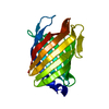 2fr2S S: Starting model for refinement C: citing same article ( |
|---|---|
| Similar structure data |


-Links
 PDBj
PDBj


- Assembly
| Deposited unit | 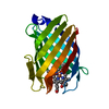
| ||||||||
|---|---|---|---|---|---|---|---|---|---|
| 1 |
| ||||||||
| Unit cell |
|
-Components
| #1: Protein | Mass: 18940.438 Da / Num. of mol.: 1 Source method: isolated from a genetically manipulated source Source: (gene. exp.) Mycobacterium tuberculosis (bacteria)Production host: Variant (production host): BL12 / References: UniProt: A0A0E8TXJ8, UniProt: P9WFG7*PLUS |
|---|---|
| #2: Chemical | ChemComp-HEM /   Mass: 616.487 Da / Num. of mol.: 1 / Source method: obtained synthetically / Formula: C34H32FeN4O4 Mass: 616.487 Da / Num. of mol.: 1 / Source method: obtained synthetically / Formula: C34H32FeN4O4 |
| #3: Chemical | ChemComp-CYN /   Mass: 26.017 Da / Num. of mol.: 1 / Source method: obtained synthetically / Formula: CN Mass: 26.017 Da / Num. of mol.: 1 / Source method: obtained synthetically / Formula: CN |
| #4: Water | ChemComp-HOH /  Mass: 18.015 Da / Num. of mol.: 77 / Source method: isolated from a natural source / Formula: H2O Mass: 18.015 Da / Num. of mol.: 77 / Source method: isolated from a natural source / Formula: H2O |
| Has protein modification | Y |
-Experimental details
-Experiment
| Experiment | Method: X-RAY DIFFRACTION / Number of used crystals: 1 |
|---|
- Sample preparation
| Crystal | Density Matthews: 2.4 Å3/Da / Density % sol: 48.83 % |
|---|---|
| Crystal grow | Temperature: 294 K / Method: vapor diffusion, hanging drop / pH: 8.5 Details: 0.02 M NiCl2, 0.1 M Tris, PEG MM 2k 20%, 0.1 M potassium cyanide |
-Data collection
| Diffraction | Mean temperature: 100 K / Serial crystal experiment: N |
|---|---|
| Diffraction source | Source: SYNCHROTRON / Site: ESRF  / Beamline: ID23-2 / Wavelength: 0.9 Å / Beamline: ID23-2 / Wavelength: 0.9 Å |
| Detector | Type: DECTRIS PILATUS3 X 2M / Detector: PIXEL / Date: Jun 14, 2018 |
| Radiation | Protocol: SINGLE WAVELENGTH / Monochromatic (M) / Laue (L): M / Scattering type: x-ray |
| Radiation wavelength | Wavelength: 0.9 Å / Relative weight: 1 |
| Reflection | Resolution: 1.6→47 Å / Num. obs: 24489 / % possible obs: 100 % / Redundancy: 8 % / Net I/σ(I): 7.6 |
| Reflection shell | Resolution: 1.6→1.69 Å / Redundancy: 8.1 % / Mean I/σ(I) obs: 2.9 / Num. unique obs: 3536 / % possible all: 100 |
- Processing
| Software |
| ||||||||||||||||||||||||||||||||||||||||||||||||||||||||||||||||||||||||||||||||||||||||||||||||||||||||||||||||||||||||||||||||||||||||||||||||||||||||||||||||||||||||||||||||||||||
|---|---|---|---|---|---|---|---|---|---|---|---|---|---|---|---|---|---|---|---|---|---|---|---|---|---|---|---|---|---|---|---|---|---|---|---|---|---|---|---|---|---|---|---|---|---|---|---|---|---|---|---|---|---|---|---|---|---|---|---|---|---|---|---|---|---|---|---|---|---|---|---|---|---|---|---|---|---|---|---|---|---|---|---|---|---|---|---|---|---|---|---|---|---|---|---|---|---|---|---|---|---|---|---|---|---|---|---|---|---|---|---|---|---|---|---|---|---|---|---|---|---|---|---|---|---|---|---|---|---|---|---|---|---|---|---|---|---|---|---|---|---|---|---|---|---|---|---|---|---|---|---|---|---|---|---|---|---|---|---|---|---|---|---|---|---|---|---|---|---|---|---|---|---|---|---|---|---|---|---|---|---|---|---|
| Refinement | Method to determine structure: MOLECULAR REPLACEMENT Starting model: 2fr2 Resolution: 1.6→47 Å / Cor.coef. Fo:Fc: 0.955 / Cor.coef. Fo:Fc free: 0.945 / SU B: 1.565 / SU ML: 0.054 / Cross valid method: THROUGHOUT / ESU R: 0.08 / ESU R Free: 0.08 / Details: HYDROGENS HAVE BEEN ADDED IN THE RIDING POSITIONS
| ||||||||||||||||||||||||||||||||||||||||||||||||||||||||||||||||||||||||||||||||||||||||||||||||||||||||||||||||||||||||||||||||||||||||||||||||||||||||||||||||||||||||||||||||||||||
| Solvent computation | Ion probe radii: 0.8 Å / Shrinkage radii: 0.8 Å / VDW probe radii: 1.2 Å | ||||||||||||||||||||||||||||||||||||||||||||||||||||||||||||||||||||||||||||||||||||||||||||||||||||||||||||||||||||||||||||||||||||||||||||||||||||||||||||||||||||||||||||||||||||||
| Displacement parameters | Biso mean: 12.119 Å2
| ||||||||||||||||||||||||||||||||||||||||||||||||||||||||||||||||||||||||||||||||||||||||||||||||||||||||||||||||||||||||||||||||||||||||||||||||||||||||||||||||||||||||||||||||||||||
| Refinement step | Cycle: 1 / Resolution: 1.6→47 Å
| ||||||||||||||||||||||||||||||||||||||||||||||||||||||||||||||||||||||||||||||||||||||||||||||||||||||||||||||||||||||||||||||||||||||||||||||||||||||||||||||||||||||||||||||||||||||
| Refine LS restraints |
|