 Movie
Movie Controller

[English] 日本語
 Yorodumi
- PDB-6qur: Mapping the allosteric communication network of aminodeoxychorism... -
+ Open data

- Basic information
| Entry | Database: PDB / ID: 6qur | ||||||
|---|---|---|---|---|---|---|---|
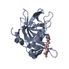
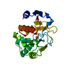
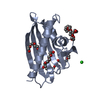
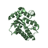
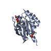
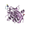
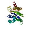
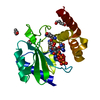
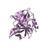
| Title | Mapping the allosteric communication network of aminodeoxychorismate synthase | ||||||
 Components Components | Glutaminase | ||||||
 Keywords Keywords | TRANSFERASE / allostery / ancestral sequence reconstruction / catalytic triad / glutamine amidotransferases | ||||||
| Function / homology | Class I glutamine amidotransferase (GATase) domain / Rossmann fold / 3-Layer(aba) Sandwich / Alpha Beta Function and homology information Function and homology information | ||||||
| Biological species | synthetic construct (others) | ||||||
| Method |  X-RAY DIFFRACTION / SYNCHROTRON / MOLECULAR REPLACEMENT / Resolution: 1.792 Å X-RAY DIFFRACTION / SYNCHROTRON / MOLECULAR REPLACEMENT / Resolution: 1.792 Å | ||||||
 Authors Authors | Semmelmann, F. / Straub, K. / Rajendran, C. / Nazet, J. | ||||||
| Funding support |  Germany, 1items Germany, 1items
| ||||||
 Citation Citation | Journal: J.Mol.Biol. / Year: 2019 Title: Mapping the Allosteric Communication Network of Aminodeoxychorismate Synthase. Authors: Semmelmann, F. / Straub, K. / Nazet, J. / Rajendran, C. / Merkl, R. / Sterner, R. | ||||||
| History |
|
- Structure visualization
| Structure viewer | Molecule: MolmilJmol/JSmol |
|---|
- Downloads & links
-Download
| PDBx/mmCIF format | 6qur.cif.gz | 54.2 KB | Display | PDBx/mmCIF format |
|---|---|---|---|---|
| PDB format | pdb6qur.ent.gz | 37.3 KB | Display | PDB format |
| PDBx/mmJSON format | 6qur.json.gz | Tree view | PDBx/mmJSON format | |
| Others |  Other downloads Other downloads |
-Validation report
| Arichive directory | https://data.pdbj.org/pub/pdb/validation_reports/qu/6qurftp://data.pdbj.org/pub/pdb/validation_reports/qu/6qur | HTTPS FTP |
|---|
-Related structure data
| Similar structure data |
|---|

-Links
 PDBj
PDBj- Assembly
| Deposited unit | 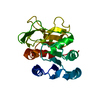
| ||||||||
|---|---|---|---|---|---|---|---|---|---|
| 1 |
| ||||||||
| Unit cell |
|
-Components
| #1: Protein | Mass: 22321.461 Da / Num. of mol.: 1 Source method: isolated from a genetically manipulated source Source: (gene. exp.) synthetic construct (others) / Production host:  |
|---|---|
| #2: Water | ChemComp-HOH /  Mass: 18.015 Da / Num. of mol.: 113 / Source method: isolated from a natural source / Formula: H2O Mass: 18.015 Da / Num. of mol.: 113 / Source method: isolated from a natural source / Formula: H2O |
| Has protein modification | Y |
-Experimental details
-Experiment
| Experiment | Method: X-RAY DIFFRACTION / Number of used crystals: 1 |
|---|
- Sample preparation
| Crystal | Density Matthews: 2.24 Å3/Da / Density % sol: 44.98 % |
|---|---|
| Crystal grow | Temperature: 291 K / Method: vapor diffusion / Details: PEG |
-Data collection
| Diffraction | Mean temperature: 100 K / Serial crystal experiment: N |
|---|---|
| Diffraction source | Source: SYNCHROTRON / Site: SLS  / Beamline: X06DA / Wavelength: 1 Å / Beamline: X06DA / Wavelength: 1 Å |
| Detector | Type: DECTRIS EIGER X 16M / Detector: PIXEL / Date: Jul 24, 2017 |
| Radiation | Protocol: SINGLE WAVELENGTH / Monochromatic (M) / Laue (L): M / Scattering type: x-ray |
| Radiation wavelength | Wavelength: 1 Å / Relative weight: 1 |
| Reflection | Resolution: 1.792→39.73 Å / Num. obs: 19372 / % possible obs: 99.87 % / Redundancy: 13 % / Biso Wilson estimate: 33.96 Å2 / CC1/2: 1 / Rmerge(I) obs: 0.06472 / Rpim(I) all: 0.01866 / Rrim(I) all: 0.06743 / Net I/σ(I): 23.52 |
| Reflection shell | Resolution: 1.79→1.856 Å / Redundancy: 11.8 % / Rmerge(I) obs: 1.935 / Mean I/σ(I) obs: 1.27 / Num. unique obs: 1880 / CC1/2: 0.866 / Rpim(I) all: 0.5803 / Rrim(I) all: 2.023 / % possible all: 99.16 |
- Processing
| Software |
| ||||||||||||||||||||||||||||||||||||||||||||||||||||||||
|---|---|---|---|---|---|---|---|---|---|---|---|---|---|---|---|---|---|---|---|---|---|---|---|---|---|---|---|---|---|---|---|---|---|---|---|---|---|---|---|---|---|---|---|---|---|---|---|---|---|---|---|---|---|---|---|---|---|
| Refinement | Method to determine structure: MOLECULAR REPLACEMENT / Resolution: 1.792→39.73 Å / SU ML: 0.25 / Cross valid method: FREE R-VALUE / σ(F): 1.35 / Phase error: 28.89
| ||||||||||||||||||||||||||||||||||||||||||||||||||||||||
| Solvent computation | Shrinkage radii: 0.9 Å / VDW probe radii: 1.11 Å | ||||||||||||||||||||||||||||||||||||||||||||||||||||||||
| Refinement step | Cycle: LAST / Resolution: 1.792→39.73 Å
| ||||||||||||||||||||||||||||||||||||||||||||||||||||||||
| Refine LS restraints |
| ||||||||||||||||||||||||||||||||||||||||||||||||||||||||
| LS refinement shell |
|
