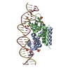
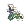
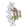
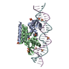
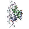
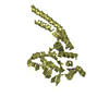
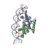
A: hsRosR-DNA binding protein B: hsRosR-DNA binding protein C: hsRosR-DNA binding protein D: hsRosR-DNA binding protein E: DNA (28-MER) F: DNA (28-MER) G: DNA (28-MER) H: DNA (28-MER) hetero molecules
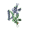
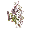
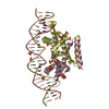
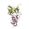
A: hsRosR-DNA binding protein B: hsRosR-DNA binding protein E: DNA (28-MER) F: DNA (28-MER) hetero molecules
defined by author
Evidence: SAXS, SAXS results indicating that one protein dimer bound a single sequence., gel filtration, The elution volume of the complex indicated that hsRosR binds one DNA molecule as a dimer., ...Evidence: SAXS, SAXS results indicating that one protein dimer bound a single sequence., gel filtration, The elution volume of the complex indicated that hsRosR binds one DNA molecule as a dimer., isothermal titration calorimetry, The binding stoichiometry was approximately one, indicating that one protein dimer bound a single sequence.
Mass: 18.015 Da / Num. of mol.: 51 / Source method: isolated from a natural source / Formula: H2O
-
Experimental details
-
Experiment
Experiment
Method: X-RAY DIFFRACTION / Number of used crystals: 1
-
Sample preparation
Crystal
Density Matthews: 4.03 Å3/Da / Density % sol: 69.45 %
Crystal grow
Temperature: 293.15 K / Method: vapor diffusion, sitting drop / pH: 7 Details: The complex crystallized from a buffer containing 20 mM HEPES, pH 7, 2 M KCl and 0.02% azide, DNA/hsRosR ratio = 1.83. 2.96 M (NH4)2SO4, 30 mM MnCl2 and 0.02% azide.
-
Data collection
Diffraction
Mean temperature: 100 K / Serial crystal experiment: N
Protocol: SINGLE WAVELENGTH / Monochromatic (M) / Laue (L): M / Scattering type: x-ray
Radiation wavelength
Wavelength: 0.976 Å / Relative weight: 1
Reflection
Resolution: 2.56→45.79 Å / Num. obs: 22780 / % possible obs: 84.4 % / Redundancy: 3.3 % / Rmerge(I) obs: 0.089 / Net I/σ(I): 7.4
Reflection shell
Resolution: 2.56→2.839 Å
-
Processing
Software
Name
Version
Classification
PHENIX
(1.14_3260: ???)
refinement
XDS
datareduction
Aimless
datascaling
STARANISO
datascaling
PHASER
phasing
Refinement
Method to determine structure: MOLECULAR REPLACEMENT Starting model: The starting model for molecular replacement was VNG0258H/RosR (from KCl; PDB code 6FDH) and a DNA model derived from Mycobacterium tuberculosis MosR (PDB code 4FX4) but with the SG sequence.
 Movie
Movie Controller


 Open data
 Basic information
 Components
 Keywords
 Function and homology information
 Halobacterium salinarum (Halophile)
 X-RAY DIFFRACTION /
  Authors
 Israel, 1items
  Citation
 Structure visualization
 Downloads & links
 Other downloads


 PDBj
PDBj


 Assembly


 Mass: 54.938 Da / Num. of mol.: 4 / Source method: obtained synthetically / Formula: Mn
 Mass: 96.063 Da / Num. of mol.: 13 / Source method: obtained synthetically / Formula: SO4
 Sample preparation
 / Beamline: ID29 / Wavelength: 0.976 Å
 Processing