 Movie
Movie Controller

[English] 日本語
 Yorodumi
- PDB-6qtv: Crystal structure of an Arabidopsis WD40 domain in complex with a... -
+ Open data

- Basic information
| Entry | Database: PDB / ID: 6qtv | ||||||||||||
|---|---|---|---|---|---|---|---|---|---|---|---|---|---|
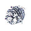
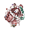
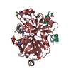
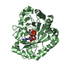
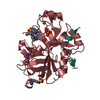
| Title | Crystal structure of an Arabidopsis WD40 domain in complex with an atypical bHLH transcription factor | ||||||||||||
 Components Components |
| ||||||||||||
 Keywords Keywords | PLANT PROTEIN / Complex | ||||||||||||
| Function / homology |  Function and homology information Function and homology informationanthocyanin-containing compound metabolic process / shade avoidance / positive regulation of flavonoid biosynthetic process / skotomorphogenesis / photoperiodism, flowering / red, far-red light phototransduction / blue light signaling pathway / response to far red light / nuclear ubiquitin ligase complex / regulation of stomatal movement ...anthocyanin-containing compound metabolic process / shade avoidance / positive regulation of flavonoid biosynthetic process / skotomorphogenesis / photoperiodism, flowering / red, far-red light phototransduction / blue light signaling pathway / response to far red light / nuclear ubiquitin ligase complex / regulation of stomatal movement / photomorphogenesis / response to light intensity / entrainment of circadian clock / response to salt / response to abscisic acid / abscisic acid-activated signaling pathway / response to UV-B / Cul4-RING E3 ubiquitin ligase complex / transcription coregulator activity / RING-type E3 ubiquitin transferase / ubiquitin-protein transferase activity / ubiquitin protein ligase activity / response to heat / proteasome-mediated ubiquitin-dependent protein catabolic process / protein dimerization activity / transcription cis-regulatory region binding / nuclear body / protein ubiquitination / DNA-binding transcription factor activity / DNA repair / zinc ion binding / identical protein binding / nucleus / cytosol / cytoplasm Similarity search - Function | ||||||||||||
| Biological species |  | ||||||||||||
| Method |  X-RAY DIFFRACTION / SYNCHROTRON / MOLECULAR REPLACEMENT / Resolution: 1.31 Å X-RAY DIFFRACTION / SYNCHROTRON / MOLECULAR REPLACEMENT / Resolution: 1.31 Å | ||||||||||||
 Authors Authors | Hothorn, M. / Lau, K. | ||||||||||||
| Funding support |  Switzerland, 3items Switzerland, 3items
| ||||||||||||
 Citation Citation | Journal: Embo J. / Year: 2019 Title: Plant photoreceptors and their signaling components compete for COP1 binding via VP peptide motifs. Authors: Lau, K. / Podolec, R. / Chappuis, R. / Ulm, R. / Hothorn, M. | ||||||||||||
| History |
|
- Structure visualization
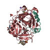
| Structure viewer | Molecule: MolmilJmol/JSmol |
|---|
- Downloads & links
-Download
| PDBx/mmCIF format | 6qtv.cif.gz | 251.1 KB | Display | PDBx/mmCIF format |
|---|---|---|---|---|
| PDB format | pdb6qtv.ent.gz | 168.1 KB | Display | PDB format |
| PDBx/mmJSON format | 6qtv.json.gz | Tree view | PDBx/mmJSON format | |
| Others |  Other downloads Other downloads |
-Validation report
| Arichive directory | https://data.pdbj.org/pub/pdb/validation_reports/qt/6qtvftp://data.pdbj.org/pub/pdb/validation_reports/qt/6qtv | HTTPS FTP |
|---|
-Related structure data
| Related structure data |  6qtoC 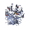 6qtqC 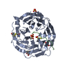 6qtrC 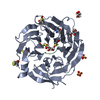 6qtsC 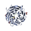 6qttC 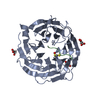 6qtuC 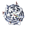 6qtwC 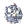 6qtxC 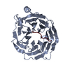 5igoS S: Starting model for refinement C: citing same article ( |
|---|---|
| Similar structure data |


-Links
 PDBj
PDBj


- Assembly
| Deposited unit | 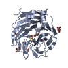
| ||||||||||||
|---|---|---|---|---|---|---|---|---|---|---|---|---|---|
| 1 |
| ||||||||||||
| Unit cell |
|
-Components
| #1: Protein | Mass: 37204.645 Da / Num. of mol.: 1 Source method: isolated from a genetically manipulated source Source: (gene. exp.)   Spodoptera frugiperda (fall armyworm) Spodoptera frugiperda (fall armyworm)References: UniProt: P43254, RING-type E3 ubiquitin transferase | ||||||
|---|---|---|---|---|---|---|---|
| #2: Protein/peptide | Mass: 1267.515 Da / Num. of mol.: 1 / Source method: obtained synthetically / Source: (synth.) | ||||||
| #3: Chemical |   Mass: 102.046 Da / Num. of mol.: 3 / Source method: obtained synthetically / Formula: C3H2O4 Mass: 102.046 Da / Num. of mol.: 3 / Source method: obtained synthetically / Formula: C3H2O4#4: Chemical |   Mass: 92.094 Da / Num. of mol.: 2 / Source method: obtained synthetically / Formula: C3H8O3 Mass: 92.094 Da / Num. of mol.: 2 / Source method: obtained synthetically / Formula: C3H8O3#5: Water | ChemComp-HOH / |  Mass: 18.015 Da / Num. of mol.: 263 / Source method: isolated from a natural source / Formula: H2O Mass: 18.015 Da / Num. of mol.: 263 / Source method: isolated from a natural source / Formula: H2OHas protein modification | Y | |
-Experimental details
-Experiment
| Experiment | Method: X-RAY DIFFRACTION / Number of used crystals: 1 |
|---|
- Sample preparation
| Crystal | Density Matthews: 1.96 Å3/Da / Density % sol: 37.17 % |
|---|---|
| Crystal grow | Temperature: 298.15 K / Method: vapor diffusion, hanging drop Details: 5 mg/mL of COP1 supplemented with 3 to 10 fold molar excess in peptide was mixed with two-fold (v/v) more mother liquor (1:2 ratio; protein:buffer) containing 1.25 M sodium malonate pH 7.5. |
-Data collection
| Diffraction | Mean temperature: 100 K / Serial crystal experiment: N |
|---|---|
| Diffraction source | Source: SYNCHROTRON / Site: SLS / Beamline: X06DA / Wavelength: 1.03 Å |
| Detector | Type: DECTRIS PILATUS 2M-F / Detector: PIXEL / Date: May 13, 2017 |
| Radiation | Protocol: SINGLE WAVELENGTH / Monochromatic (M) / Laue (L): M / Scattering type: x-ray |
| Radiation wavelength | Wavelength: 1.03 Å / Relative weight: 1 |
| Reflection | Resolution: 1.31→48.58 Å / Num. obs: 67056 / % possible obs: 99.76 % / Redundancy: 12.4 % / Biso Wilson estimate: 12.46 Å2 / Rrim(I) all: 0.0345 / Net I/σ(I): 16.09 |
| Reflection shell | Resolution: 1.31→1.36 Å |
- Processing
| Software |
| ||||||||||||||||||||||||||||||||||||||||||||||||||||||||||||||||||||||||||||||||||||||||||||||||||||||||||||||||||||||||||||||||||||||||||||||||||||||||||||||||||||||||
|---|---|---|---|---|---|---|---|---|---|---|---|---|---|---|---|---|---|---|---|---|---|---|---|---|---|---|---|---|---|---|---|---|---|---|---|---|---|---|---|---|---|---|---|---|---|---|---|---|---|---|---|---|---|---|---|---|---|---|---|---|---|---|---|---|---|---|---|---|---|---|---|---|---|---|---|---|---|---|---|---|---|---|---|---|---|---|---|---|---|---|---|---|---|---|---|---|---|---|---|---|---|---|---|---|---|---|---|---|---|---|---|---|---|---|---|---|---|---|---|---|---|---|---|---|---|---|---|---|---|---|---|---|---|---|---|---|---|---|---|---|---|---|---|---|---|---|---|---|---|---|---|---|---|---|---|---|---|---|---|---|---|---|---|---|---|---|---|---|---|
| Refinement | Method to determine structure: MOLECULAR REPLACEMENT Starting model: 5IGO Resolution: 1.31→48.58 Å / SU ML: 0.1241 / Cross valid method: FREE R-VALUE / σ(F): 1.34 / Phase error: 16.5521
| ||||||||||||||||||||||||||||||||||||||||||||||||||||||||||||||||||||||||||||||||||||||||||||||||||||||||||||||||||||||||||||||||||||||||||||||||||||||||||||||||||||||||
| Solvent computation | Shrinkage radii: 0.9 Å / VDW probe radii: 1.11 Å | ||||||||||||||||||||||||||||||||||||||||||||||||||||||||||||||||||||||||||||||||||||||||||||||||||||||||||||||||||||||||||||||||||||||||||||||||||||||||||||||||||||||||
| Displacement parameters | Biso mean: 19.45 Å2 | ||||||||||||||||||||||||||||||||||||||||||||||||||||||||||||||||||||||||||||||||||||||||||||||||||||||||||||||||||||||||||||||||||||||||||||||||||||||||||||||||||||||||
| Refinement step | Cycle: LAST / Resolution: 1.31→48.58 Å
| ||||||||||||||||||||||||||||||||||||||||||||||||||||||||||||||||||||||||||||||||||||||||||||||||||||||||||||||||||||||||||||||||||||||||||||||||||||||||||||||||||||||||
| Refine LS restraints |
| ||||||||||||||||||||||||||||||||||||||||||||||||||||||||||||||||||||||||||||||||||||||||||||||||||||||||||||||||||||||||||||||||||||||||||||||||||||||||||||||||||||||||
| LS refinement shell |
|
