 Movie
Movie Controller

[English] 日本語
 Yorodumi
- PDB-6qt1: Radiation damage study on a 16mer DNA segment, structure at 0.48 ... -
+ Open data

- Basic information
| Entry | Database: PDB / ID: 6qt1 | |||||||||||||||||||||||||||||||
|---|---|---|---|---|---|---|---|---|---|---|---|---|---|---|---|---|---|---|---|---|---|---|---|---|---|---|---|---|---|---|---|---|
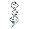
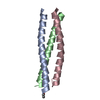
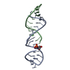
| Title | Radiation damage study on a 16mer DNA segment, structure at 0.48 MGy dose | |||||||||||||||||||||||||||||||
 Components Components | DNA (5'-D(* Keywords KeywordsDNA / radiation damage / global damage / specific damage | Function / homology | DNA / DNA (> 10) |  Function and homology information Function and homology informationBiological species | synthetic construct (others) | Method |  X-RAY DIFFRACTION / SYNCHROTRON / MOLECULAR REPLACEMENT / molecular replacement / Resolution: 1.8 Å X-RAY DIFFRACTION / SYNCHROTRON / MOLECULAR REPLACEMENT / molecular replacement / Resolution: 1.8 Å  Authors AuthorsBugris, V. / Harmat, V. / Ferenc, G. / Brockhauser, S. / Carmichael, I. / Garman, E.F. | Funding support | |  Germany, Germany,  Hungary, 2items Hungary, 2items
 CitationJournal: J.Synchrotron Radiat. / Year: 2019 CitationJournal: J.Synchrotron Radiat. / Year: 2019Title: Radiation-damage investigation of a DNA 16-mer. Authors: Bugris, V. / Harmat, V. / Ferenc, G. / Brockhauser, S. / Carmichael, I. / Garman, E.F. History |
|
- Structure visualization
| Structure viewer | Molecule: MolmilJmol/JSmol |
|---|
- Downloads & links
-Download
| PDBx/mmCIF format | 6qt1.cif.gz | 32.4 KB | Display | PDBx/mmCIF format |
|---|---|---|---|---|
| PDB format | pdb6qt1.ent.gz | 21.4 KB | Display | PDB format |
| PDBx/mmJSON format | 6qt1.json.gz | Tree view | PDBx/mmJSON format | |
| Others |  Other downloads Other downloads |
-Validation report
| Arichive directory | https://data.pdbj.org/pub/pdb/validation_reports/qt/6qt1ftp://data.pdbj.org/pub/pdb/validation_reports/qt/6qt1 | HTTPS FTP |
|---|
-Related structure data
| Related structure data |  6qt2C  6qt3C  6qt4C  6qt5C  6qt6C  1sgsS  6qt7 C: citing same article ( S: Starting model for refinement |
|---|---|
| Similar structure data |


-Links
 PDBj
PDBj


- Assembly
| Deposited unit | 
| |||||||||||||||
|---|---|---|---|---|---|---|---|---|---|---|---|---|---|---|---|---|
| 1 | 
| |||||||||||||||
| Unit cell |
| |||||||||||||||
| Components on special symmetry positions |
|
-Components
| #1: DNA chain | Mass: 4898.191 Da / Num. of mol.: 1 / Source method: obtained synthetically Details: "Self complementary model DNA sequence derived from kB33 DNA segment. The present sequence is one nucleotide shorter than that of PDB structure 1SGS. Source: (synth.) synthetic construct (others) | ||
|---|---|---|---|
| #2: Chemical | ChemComp-CA /   Mass: 40.078 Da / Num. of mol.: 6 / Source method: obtained synthetically / Formula: Ca Mass: 40.078 Da / Num. of mol.: 6 / Source method: obtained synthetically / Formula: Ca#3: Water | ChemComp-HOH / |  Mass: 18.015 Da / Num. of mol.: 38 / Source method: isolated from a natural source / Formula: H2O Mass: 18.015 Da / Num. of mol.: 38 / Source method: isolated from a natural source / Formula: H2O |
-Experimental details
-Experiment
| Experiment | Method: X-RAY DIFFRACTION / Number of used crystals: 1 |
|---|
- Sample preparation
| Crystal | Density Matthews: 2.16 Å3/Da / Density % sol: 42.97 % |
|---|---|
| Crystal grow | Temperature: 292 K / Method: vapor diffusion, hanging drop / pH: 8.6 Details: 2 microliter 1.5 mM DNA solution (5mM HEPES pH 6.6) plus 2 microliter 10 mM HEPES pH 6.6 plus 4 microliter reservoir solution, against a reservoir of 1 ml 34% PEG200, 600 mM CaCl2 and 10 mM HEPES pH 8.6. PH range: 6.6-8.6 |
-Data collection
| Diffraction | Mean temperature: 100 K / Serial crystal experiment: N | |||||||||||||||||||||
|---|---|---|---|---|---|---|---|---|---|---|---|---|---|---|---|---|---|---|---|---|---|---|
| Diffraction source | Source: SYNCHROTRON / Site: Diamond  / Beamline: I24 / Wavelength: 0.9686 Å / Beamline: I24 / Wavelength: 0.9686 Å | |||||||||||||||||||||
| Detector | Type: DECTRIS PILATUS3 6M / Detector: PIXEL / Date: Oct 22, 2018 | |||||||||||||||||||||
| Radiation | Protocol: SINGLE WAVELENGTH / Monochromatic (M) / Laue (L): M / Scattering type: x-ray | |||||||||||||||||||||
| Radiation wavelength | Wavelength: 0.9686 Å / Relative weight: 1 | |||||||||||||||||||||
| Reflection | Resolution: 1.8→31.29 Å / Num. obs: 3861 / % possible obs: 92.1 % / Redundancy: 2.9 % / CC1/2: 0.998 / Rmerge(I) obs: 0.044 / Rpim(I) all: 0.03 / Rrim(I) all: 0.053 / Net I/σ(I): 9.4 | |||||||||||||||||||||
| Reflection shell | Diffraction-ID: 1 / Redundancy: 2.5 %
|
-Phasing
| Phasing | Method: molecular replacement |
|---|
- Processing
| Software |
| ||||||||||||||||||||||||||||||||||||||||
|---|---|---|---|---|---|---|---|---|---|---|---|---|---|---|---|---|---|---|---|---|---|---|---|---|---|---|---|---|---|---|---|---|---|---|---|---|---|---|---|---|---|
| Refinement | Method to determine structure: MOLECULAR REPLACEMENT Starting model: 1SGS Resolution: 1.8→31.29 Å / Cor.coef. Fo:Fc: 0.963 / Cor.coef. Fo:Fc free: 0.94 / SU B: 7.071 / SU ML: 0.123 / Cross valid method: THROUGHOUT / σ(F): 0 / ESU R: 0.171 / ESU R Free: 0.153 Details: HYDROGENS HAVE BEEN ADDED IN THE RIDING POSITIONS U VALUES : WITH TLS ADDED. Water molecules found in close contacts because of two reasons. 1) Being a part of the coordination sphere of ...Details: HYDROGENS HAVE BEEN ADDED IN THE RIDING POSITIONS U VALUES : WITH TLS ADDED. Water molecules found in close contacts because of two reasons. 1) Being a part of the coordination sphere of Ca2+ ions, possibly in different configurations, could not be fully resolved. 2) Some water molecules are in contact with the phosphate moieties of the DNA, with some contact angles close to 90 degrees, suggesting these may be Mg2+ or Na+ ions - these were modelled as water molecules, because there were no sodium or magnesium salts present during purification and crystallization.
| ||||||||||||||||||||||||||||||||||||||||
| Solvent computation | Ion probe radii: 0.8 Å / Shrinkage radii: 0.8 Å / VDW probe radii: 1.2 Å | ||||||||||||||||||||||||||||||||||||||||
| Displacement parameters | Biso max: 101.25 Å2 / Biso mean: 49.345 Å2 / Biso min: 22.56 Å2
| ||||||||||||||||||||||||||||||||||||||||
| Refinement step | Cycle: final / Resolution: 1.8→31.29 Å
| ||||||||||||||||||||||||||||||||||||||||
| Refine LS restraints |
| ||||||||||||||||||||||||||||||||||||||||
| LS refinement shell | Resolution: 1.801→1.847 Å / Rfactor Rfree error: 0 / Total num. of bins used: 20
| ||||||||||||||||||||||||||||||||||||||||
| Refinement TLS params. | Method: refined / Origin x: -10.8379 Å / Origin y: -20.1414 Å / Origin z: 54.1618 Å
|
