 Movie
Movie Controller


+ Open data

- Basic information
| Entry | Database: PDB / ID: 1sgs | ||||||
|---|---|---|---|---|---|---|---|
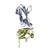
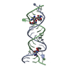
| Title | Crystal structure of a free kB DNA | ||||||
 Components Components | kB DNA | ||||||
 Keywords Keywords | DNA / free kB DNA / Calcium binding / NF-kB binding | ||||||
| Function / homology | DNA / DNA (> 10) Function and homology information Function and homology information | ||||||
| Method |  X-RAY DIFFRACTION / MOLECULAR REPLACEMENT / Resolution: 1.6 Å X-RAY DIFFRACTION / MOLECULAR REPLACEMENT / Resolution: 1.6 Å | ||||||
 Authors Authors | Huang, D.B. / Phelps, C.B. / Fusco, A.J. / Ghosh, G. | ||||||
 Citation Citation | Journal: J.Mol.Biol. / Year: 2005 Title: Crystal structure of a free kappaB DNA: insights into DNA recognition by transcription factor NF-kappaB. Authors: Huang, D.B. / Phelps, C.B. / Fusco, A.J. / Ghosh, G. | ||||||
| History |
|
- Structure visualization
| Structure viewer | Molecule: MolmilJmol/JSmol |
|---|
- Downloads & links
-Download
| PDBx/mmCIF format | 1sgs.cif.gz | 24.5 KB | Display | PDBx/mmCIF format |
|---|---|---|---|---|
| PDB format | pdb1sgs.ent.gz | 15 KB | Display | PDB format |
| PDBx/mmJSON format | 1sgs.json.gz | Tree view | PDBx/mmJSON format | |
| Others |  Other downloads Other downloads |
-Validation report
| Arichive directory | https://data.pdbj.org/pub/pdb/validation_reports/sg/1sgsftp://data.pdbj.org/pub/pdb/validation_reports/sg/1sgs | HTTPS FTP |
|---|
-Related structure data
| Related structure data |  1ramS S: Starting model for refinement |
|---|---|
| Similar structure data |


-Links
 PDBj
PDBj


- Assembly
| Deposited unit | 
| |||||||||||||||||||||
|---|---|---|---|---|---|---|---|---|---|---|---|---|---|---|---|---|---|---|---|---|---|---|
| 1 | 
| |||||||||||||||||||||
| Unit cell |
| |||||||||||||||||||||
| Components on special symmetry positions |
| |||||||||||||||||||||
| Details | The second part of the biological assembly is generated by a two fold axis in R32 space group |
-Components
| #1: DNA chain | Mass: 5187.373 Da / Num. of mol.: 1 / Source method: obtained synthetically Details: The protein was chemically synthesized. The sequence of the protein naturally occurs in Homo sapiens (human). | ||
|---|---|---|---|
| #2: Chemical | ChemComp-CA /   Mass: 40.078 Da / Num. of mol.: 6 / Source method: obtained synthetically / Formula: Ca Mass: 40.078 Da / Num. of mol.: 6 / Source method: obtained synthetically / Formula: Ca#3: Water | ChemComp-HOH / |  Mass: 18.015 Da / Num. of mol.: 107 / Source method: isolated from a natural source / Formula: H2O Mass: 18.015 Da / Num. of mol.: 107 / Source method: isolated from a natural source / Formula: H2O |
-Experimental details
-Experiment
| Experiment | Method: X-RAY DIFFRACTION / Number of used crystals: 1 |
|---|
- Sample preparation
| Crystal | Density Matthews: 2.23 Å3/Da / Density % sol: 44.9 % | |||||||||||||||||||||||||||||||||||||||||||||||||
|---|---|---|---|---|---|---|---|---|---|---|---|---|---|---|---|---|---|---|---|---|---|---|---|---|---|---|---|---|---|---|---|---|---|---|---|---|---|---|---|---|---|---|---|---|---|---|---|---|---|---|
| Crystal grow | Temperature: 291 K / Method: vapor diffusion, hanging drop / pH: 7.5 Details: PEG 3350, BOG, CaCl2, pH 7.5, VAPOR DIFFUSION, HANGING DROP, temperature 291K | |||||||||||||||||||||||||||||||||||||||||||||||||
| Crystal grow | *PLUS Temperature: 18 ℃ / Method: vapor diffusion, hanging drop | |||||||||||||||||||||||||||||||||||||||||||||||||
| Components of the solutions | *PLUS
|
-Data collection
| Diffraction | Mean temperature: 105 K |
|---|---|
| Diffraction source | Source: ROTATING ANODE / Type: RIGAKU RU200 / Wavelength: 1.5418 Å |
| Detector | Type: MARRESEARCH / Detector: AREA DETECTOR / Date: Jul 1, 2003 / Details: mirrors |
| Radiation | Monochromator: osmic mirror / Protocol: SINGLE WAVELENGTH / Monochromatic (M) / Laue (L): M / Scattering type: x-ray |
| Radiation wavelength | Wavelength: 1.5418 Å / Relative weight: 1 |
| Reflection | Resolution: 1.6→30 Å / Num. all: 6411 / Num. obs: 6341 / % possible obs: 98.8 % / Observed criterion σ(F): 0 / Observed criterion σ(I): 0 / Redundancy: 37 % / Biso Wilson estimate: 16.5 Å2 / Rmerge(I) obs: 0.39 / Net I/σ(I): 29.4 |
| Reflection shell | Resolution: 1.6→1.66 Å / Rmerge(I) obs: 0.26 / Mean I/σ(I) obs: 12 / % possible all: 91.3 |
| Reflection | *PLUS % possible obs: 99 % / Num. measured all: 242967 / Rmerge(I) obs: 0.039 |
| Reflection shell | *PLUS % possible obs: 91 % / Rmerge(I) obs: 0.256 / Mean I/σ(I) obs: 12 |
- Processing
| Software |
| |||||||||||||||||||||||||
|---|---|---|---|---|---|---|---|---|---|---|---|---|---|---|---|---|---|---|---|---|---|---|---|---|---|---|
| Refinement | Method to determine structure: MOLECULAR REPLACEMENT Starting model: PDB entry 1RAM Resolution: 1.6→14.37 Å / Rfactor Rfree error: 0.009 / Data cutoff high absF: 1003948.67 / Data cutoff low absF: 0 / Isotropic thermal model: RESTRAINED / Cross valid method: THROUGHOUT / σ(F): 0
| |||||||||||||||||||||||||
| Solvent computation | Solvent model: FLAT MODEL / Bsol: 43.4827 Å2 / ksol: 0.380301 e/Å3 | |||||||||||||||||||||||||
| Displacement parameters | Biso mean: 24.9 Å2
| |||||||||||||||||||||||||
| Refine analyze |
| |||||||||||||||||||||||||
| Refinement step | Cycle: LAST / Resolution: 1.6→14.37 Å
| |||||||||||||||||||||||||
| Refine LS restraints |
| |||||||||||||||||||||||||
| LS refinement shell | Resolution: 1.6→1.7 Å / Rfactor Rfree error: 0.039 / Total num. of bins used: 6
| |||||||||||||||||||||||||
| Xplor file |
| |||||||||||||||||||||||||
| Refinement | *PLUS Lowest resolution: 30 Å / Num. reflection obs: 6340 / % reflection Rfree: 10 % / Rfactor Rfree: 0.215 | |||||||||||||||||||||||||
| Solvent computation | *PLUS | |||||||||||||||||||||||||
| Displacement parameters | *PLUS | |||||||||||||||||||||||||
| Refine LS restraints | *PLUS
|
