[English] 日本語
 Yorodumi
- PDB-6qpq: The structure of the cohesin head module elucidates the mechanism... -
+ Open data
- Basic information
| Entry | Database: PDB / ID: 6qpq | ||||||
|---|---|---|---|---|---|---|---|
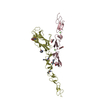
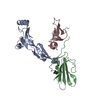
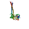
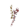
| Title | The structure of the cohesin head module elucidates the mechanism of ring opening | ||||||
 Components Components |
| ||||||
 Keywords Keywords | CELL CYCLE / Cohesin / cell division / genome regulation / sister chromatid cohesion / SMC / kleisin | ||||||
| Function / homology |  Function and homology information Function and homology informationEstablishment of Sister Chromatid Cohesion / Resolution of Sister Chromatid Cohesion / cohesin complex / mitotic cohesin complex / SUMOylation of DNA damage response and repair proteins / replication-born double-strand break repair via sister chromatid exchange / establishment of mitotic sister chromatid cohesion / mitotic chromosome condensation / sister chromatid cohesion / mitotic sister chromatid cohesion ...Establishment of Sister Chromatid Cohesion / Resolution of Sister Chromatid Cohesion / cohesin complex / mitotic cohesin complex / SUMOylation of DNA damage response and repair proteins / replication-born double-strand break repair via sister chromatid exchange / establishment of mitotic sister chromatid cohesion / mitotic chromosome condensation / sister chromatid cohesion / mitotic sister chromatid cohesion / protein acetylation / chromosome, centromeric region / condensed nuclear chromosome / G2/M transition of mitotic cell cycle / double-strand break repair / cell division / apoptotic process / DNA damage response / chromatin binding / protein kinase binding / ATP hydrolysis activity / mitochondrion / DNA binding / ATP binding / nucleus Similarity search - Function | ||||||
| Biological species |  Chaetomium thermophilum var. thermophilum DSM 1495 (fungus) Chaetomium thermophilum var. thermophilum DSM 1495 (fungus) | ||||||
| Method |  X-RAY DIFFRACTION / X-RAY DIFFRACTION /  SYNCHROTRON / SYNCHROTRON /  MOLECULAR REPLACEMENT / Resolution: 2.1 Å MOLECULAR REPLACEMENT / Resolution: 2.1 Å | ||||||
 Authors Authors | Li, Y. / Muir, K.W. / Panne, D. | ||||||
 Citation Citation |  Journal: Nat Struct Mol Biol / Year: 2020 Journal: Nat Struct Mol Biol / Year: 2020Title: The structure of the cohesin ATPase elucidates the mechanism of SMC-kleisin ring opening. Authors: Kyle W Muir / Yan Li / Felix Weis / Daniel Panne /    Abstract: Genome regulation requires control of chromosome organization by SMC-kleisin complexes. The cohesin complex contains the Smc1 and Smc3 subunits that associate with the kleisin Scc1 to form a ring- ...Genome regulation requires control of chromosome organization by SMC-kleisin complexes. The cohesin complex contains the Smc1 and Smc3 subunits that associate with the kleisin Scc1 to form a ring-shaped complex that can topologically engage chromatin to regulate chromatin structure. Release from chromatin involves opening of the ring at the Smc3-Scc1 interface in a reaction that is controlled by acetylation and engagement of the Smc ATPase head domains. To understand the underlying molecular mechanisms, we have determined the 3.2-Å resolution cryo-electron microscopy structure of the ATPγS-bound, heterotrimeric cohesin ATPase head module and the 2.1-Å resolution crystal structure of a nucleotide-free Smc1-Scc1 subcomplex from Saccharomyces cerevisiae and Chaetomium thermophilium. We found that ATP-binding and Smc1-Smc3 heterodimerization promote conformational changes within the ATPase that are transmitted to the Smc coiled-coil domains. Remodeling of the coiled-coil domain of Smc3 abrogates the binding surface for Scc1, thus leading to ring opening at the Smc3-Scc1 interface. | ||||||
| History |
|
- Structure visualization
| Structure viewer | Molecule:  Molmil Molmil Jmol/JSmol Jmol/JSmol |
|---|
- Downloads & links
- Download
| PDBx/mmCIF format |  6qpq.cif.gz 6qpq.cif.gz | 481 KB | Display |  PDBx/mmCIF format PDBx/mmCIF format |
|---|---|---|---|---|
| PDB format |  pdb6qpq.ent.gz pdb6qpq.ent.gz | 313.9 KB | Display |  PDB format PDB format |
| PDBx/mmJSON format |  6qpq.json.gz 6qpq.json.gz | Tree view |  PDBx/mmJSON format PDBx/mmJSON format | |
| Others |  Other downloads Other downloads |
-Validation report
| Summary document |  6qpq_validation.pdf.gz 6qpq_validation.pdf.gz | 432.9 KB | Display |  wwPDB validaton report wwPDB validaton report |
|---|---|---|---|---|
| Full document |  6qpq_full_validation.pdf.gz 6qpq_full_validation.pdf.gz | 436 KB | Display | |
| Data in XML |  6qpq_validation.xml.gz 6qpq_validation.xml.gz | 19.1 KB | Display | |
| Data in CIF |  6qpq_validation.cif.gz 6qpq_validation.cif.gz | 30.3 KB | Display | |
| Arichive directory |  https://data.pdbj.org/pub/pdb/validation_reports/qp/6qpq https://data.pdbj.org/pub/pdb/validation_reports/qp/6qpq ftp://data.pdbj.org/pub/pdb/validation_reports/qp/6qpq ftp://data.pdbj.org/pub/pdb/validation_reports/qp/6qpq | HTTPS FTP |
-Related structure data
- Links
Links
- Assembly
| Deposited unit | 
| ||||||||||||
|---|---|---|---|---|---|---|---|---|---|---|---|---|---|
| 1 | 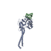
| ||||||||||||
| 2 | 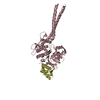
| ||||||||||||
| Unit cell |
|
- Components
| #1: Protein | Mass: 51883.062 Da / Num. of mol.: 2 Source method: isolated from a genetically manipulated source Source: (gene. exp.)  Chaetomium thermophilum var. thermophilum DSM 1495 (fungus) Chaetomium thermophilum var. thermophilum DSM 1495 (fungus)Gene: CTHT_0066330 / Production host:  #2: Protein | Mass: 63352.852 Da / Num. of mol.: 2 Source method: isolated from a genetically manipulated source Source: (gene. exp.)   #3: Water | ChemComp-HOH / | |
|---|
-Experimental details
-Experiment
| Experiment | Method:  X-RAY DIFFRACTION / Number of used crystals: 1 X-RAY DIFFRACTION / Number of used crystals: 1 |
|---|
- Sample preparation
| Crystal grow | Temperature: 277 K / Method: vapor diffusion, hanging drop / Details: 0.1 M Tris pH 8.5, 30% PEG300 |
|---|
-Data collection
| Diffraction | Mean temperature: 100 K / Serial crystal experiment: N |
|---|---|
| Diffraction source | Source:  SYNCHROTRON / Site: SYNCHROTRON / Site:  ESRF ESRF  / Beamline: MASSIF-1 / Wavelength: 0.966 Å / Beamline: MASSIF-1 / Wavelength: 0.966 Å |
| Detector | Type: DECTRIS PILATUS3 2M / Detector: PIXEL / Date: Mar 9, 2016 |
| Radiation | Protocol: SINGLE WAVELENGTH / Monochromatic (M) / Laue (L): M / Scattering type: x-ray |
| Radiation wavelength | Wavelength: 0.966 Å / Relative weight: 1 |
| Reflection | Resolution: 2.1→45.7 Å / Num. obs: 82453 / % possible obs: 98.8 % / Redundancy: 4.5 % / Net I/σ(I): 13.36 |
| Reflection shell | Resolution: 2.14→2.27 Å |
- Processing
| Software |
| ||||||||||||
|---|---|---|---|---|---|---|---|---|---|---|---|---|---|
| Refinement | Method to determine structure:  MOLECULAR REPLACEMENT / Resolution: 2.1→45.7 Å / Cross valid method: FREE R-VALUE / MOLECULAR REPLACEMENT / Resolution: 2.1→45.7 Å / Cross valid method: FREE R-VALUE /
| ||||||||||||
| Refinement step | Cycle: LAST / Resolution: 2.1→45.7 Å
|
 Movie
Movie Controller


 PDBj
PDBj
