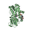
 Movie
Movie Controller

[English] 日本語
 Yorodumi
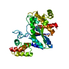
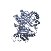
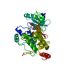
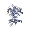
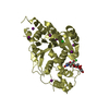
- PDB-6qmo: Death-associated Protein Kinase 1 (DAPK1) catalytic and auto-regu... -
+ Open data

- Basic information
| Entry | Database: PDB / ID: 6qmo | ||||||
|---|---|---|---|---|---|---|---|
| Title | Death-associated Protein Kinase 1 (DAPK1) catalytic and auto-regulatory domains with S289E and S308A mutations | ||||||
 Components Components | Death-associated protein kinase 1 | ||||||
 Keywords Keywords | SIGNALING PROTEIN / kinase / apoptosis / autophagy / CaMK | ||||||
| Function / homology |  Function and homology information Function and homology informationcellular response to hydroperoxide / regulation of response to tumor cell / positive regulation of autophagic cell death / DAPK1-calmodulin complex / Caspase activation via Dependence Receptors in the absence of ligand / defense response to tumor cell / calcium/calmodulin-dependent protein kinase activity / regulation of NMDA receptor activity / syntaxin-1 binding / extrinsic apoptotic signaling pathway via death domain receptors ...cellular response to hydroperoxide / regulation of response to tumor cell / positive regulation of autophagic cell death / DAPK1-calmodulin complex / Caspase activation via Dependence Receptors in the absence of ligand / defense response to tumor cell / calcium/calmodulin-dependent protein kinase activity / regulation of NMDA receptor activity / syntaxin-1 binding / extrinsic apoptotic signaling pathway via death domain receptors / positive regulation of autophagy / regulation of autophagy / apoptotic signaling pathway / cellular response to type II interferon / protein autophosphorylation / actin cytoskeleton / regulation of apoptotic process / protein phosphorylation / protein kinase activity / calmodulin binding / non-specific serine/threonine protein kinase / negative regulation of translation / postsynaptic density / intracellular signal transduction / positive regulation of apoptotic process / protein serine kinase activity / protein serine/threonine kinase activity / apoptotic process / negative regulation of apoptotic process / GTP binding / glutamatergic synapse / ATP binding / identical protein binding / nucleus / plasma membrane / cytoplasm Similarity search - Function | ||||||
| Biological species |  Homo sapiens (human) Homo sapiens (human) | ||||||
| Method |  X-RAY DIFFRACTION / SYNCHROTRON / MOLECULAR REPLACEMENT / Resolution: 1.87 Å X-RAY DIFFRACTION / SYNCHROTRON / MOLECULAR REPLACEMENT / Resolution: 1.87 Å | ||||||
 Authors Authors | Huart, A.-S. / Wilmanns, M. | ||||||
 Citation Citation | Journal: To Be Published Title: Molecular mechanisms behind DAPK regulation: how phosphorylation switches work Authors: Huart, A.-S. / Simon, B. / Lubner, J. / Mertens, H.D.T. / Temmerman, K. / Hoffmann, J.-E. / Svergun, D.I. / Schwartz, D. / Schultz, C. / Wilmanns, M. | ||||||
| History |
|
- Structure visualization
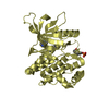
| Structure viewer | Molecule: MolmilJmol/JSmol |
|---|
- Downloads & links
-Download
| PDBx/mmCIF format | 6qmo.cif.gz | 140.2 KB | Display | PDBx/mmCIF format |
|---|---|---|---|---|
| PDB format | pdb6qmo.ent.gz | 108.9 KB | Display | PDB format |
| PDBx/mmJSON format | 6qmo.json.gz | Tree view | PDBx/mmJSON format | |
| Others |  Other downloads Other downloads |
-Validation report
| Arichive directory | https://data.pdbj.org/pub/pdb/validation_reports/qm/6qmoftp://data.pdbj.org/pub/pdb/validation_reports/qm/6qmo | HTTPS FTP |
|---|
-Related structure data
| Related structure data |  6fhaC  6fhbC  6qn4C 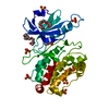 4b4lS C: citing same article ( S: Starting model for refinement |
|---|---|
| Similar structure data |

-Links
 PDBj
PDBj


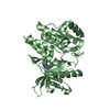
- Assembly
| Deposited unit | 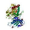
| ||||||||
|---|---|---|---|---|---|---|---|---|---|
| 1 |
| ||||||||
| Unit cell |
|
-Components
| #1: Protein | Mass: 35992.215 Da / Num. of mol.: 1 / Mutation: S289E S308A Source method: isolated from a genetically manipulated source Source: (gene. exp.) Homo sapiens (human) / Gene: DAPK1, DAPK / Production host:  References: UniProt: P53355, non-specific serine/threonine protein kinase | ||||||
|---|---|---|---|---|---|---|---|
| #2: Chemical | ChemComp-GOL /   Mass: 92.094 Da / Num. of mol.: 6 / Source method: obtained synthetically / Formula: C3H8O3 Mass: 92.094 Da / Num. of mol.: 6 / Source method: obtained synthetically / Formula: C3H8O3#3: Chemical | ChemComp-CL /   Mass: 35.453 Da / Num. of mol.: 5 / Source method: obtained synthetically / Formula: Cl Mass: 35.453 Da / Num. of mol.: 5 / Source method: obtained synthetically / Formula: Cl#4: Chemical | ChemComp-PGE / |   Mass: 150.173 Da / Num. of mol.: 1 / Source method: obtained synthetically / Formula: C6H14O4 Mass: 150.173 Da / Num. of mol.: 1 / Source method: obtained synthetically / Formula: C6H14O4#5: Water | ChemComp-HOH / |  Mass: 18.015 Da / Num. of mol.: 115 / Source method: isolated from a natural source / Formula: H2O Mass: 18.015 Da / Num. of mol.: 115 / Source method: isolated from a natural source / Formula: H2O |
-Experimental details
-Experiment
| Experiment | Method: X-RAY DIFFRACTION / Number of used crystals: 1 |
|---|
- Sample preparation
| Crystal | Density Matthews: 2.32 Å3/Da / Density % sol: 47.01 % / Description: plate needle |
|---|---|
| Crystal grow | Temperature: 292.15 K / Method: vapor diffusion, sitting drop / Details: 20% PEG 3350, 0.2M magnesium formate |
-Data collection
| Diffraction | Mean temperature: 100 K / Serial crystal experiment: N |
|---|---|
| Diffraction source | Source: SYNCHROTRON / Site: PETRA III, EMBL c/o DESY  / Beamline: P13 (MX1) / Wavelength: 1.0332 Å / Beamline: P13 (MX1) / Wavelength: 1.0332 Å |
| Detector | Type: DECTRIS PILATUS 6M-F / Detector: PIXEL / Date: Nov 8, 2016 / Details: Toroidal mirror |
| Radiation | Monochromator: Si(111) / Protocol: SINGLE WAVELENGTH / Monochromatic (M) / Laue (L): M / Scattering type: x-ray |
| Radiation wavelength | Wavelength: 1.0332 Å / Relative weight: 1 |
| Reflection | Resolution: 1.81→47.21 Å / Num. obs: 31119 / % possible obs: 98.8 % / Redundancy: 6.6 % / Rmerge(I) obs: 0.123 / Rpim(I) all: 0.051 / Rrim(I) all: 0.133 / Net I/σ(I): 9.3 |
| Reflection shell | Resolution: 1.81→1.87 Å / Redundancy: 6.6 % / Rmerge(I) obs: 1.1 / Mean I/σ(I) obs: 1.5 / CC1/2: 0.242 / Rpim(I) all: 0.457 / Rrim(I) all: 1.193 / % possible all: 97.1 |
- Processing
| Software |
| ||||||||||||||||||||||||||||||||||||||||||||||||||||||||||||||||||||||||||||||||||||||||||||||||||||
|---|---|---|---|---|---|---|---|---|---|---|---|---|---|---|---|---|---|---|---|---|---|---|---|---|---|---|---|---|---|---|---|---|---|---|---|---|---|---|---|---|---|---|---|---|---|---|---|---|---|---|---|---|---|---|---|---|---|---|---|---|---|---|---|---|---|---|---|---|---|---|---|---|---|---|---|---|---|---|---|---|---|---|---|---|---|---|---|---|---|---|---|---|---|---|---|---|---|---|---|---|---|
| Refinement | Method to determine structure: MOLECULAR REPLACEMENT Starting model: 4B4L Resolution: 1.87→42.182 Å / SU ML: 0.21 / Cross valid method: FREE R-VALUE / σ(F): 1.93 / Phase error: 22.2
| ||||||||||||||||||||||||||||||||||||||||||||||||||||||||||||||||||||||||||||||||||||||||||||||||||||
| Solvent computation | Shrinkage radii: 0.9 Å / VDW probe radii: 1.11 Å | ||||||||||||||||||||||||||||||||||||||||||||||||||||||||||||||||||||||||||||||||||||||||||||||||||||
| Refinement step | Cycle: LAST / Resolution: 1.87→42.182 Å
| ||||||||||||||||||||||||||||||||||||||||||||||||||||||||||||||||||||||||||||||||||||||||||||||||||||
| Refine LS restraints |
| ||||||||||||||||||||||||||||||||||||||||||||||||||||||||||||||||||||||||||||||||||||||||||||||||||||
| LS refinement shell |
| ||||||||||||||||||||||||||||||||||||||||||||||||||||||||||||||||||||||||||||||||||||||||||||||||||||
| Refinement TLS params. | Method: refined / Refine-ID: X-RAY DIFFRACTION
| ||||||||||||||||||||||||||||||||||||||||||||||||||||||||||||||||||||||||||||||||||||||||||||||||||||
| Refinement TLS group |
|
