 Movie
Movie Controller

[English] 日本語
 Yorodumi
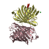
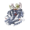
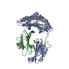
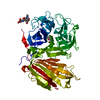
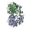
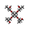
- PDB-6qmm: Crystal structure of Synecochoccus Spermidine Synthase in complex... -
+ Open data

- Basic information
| Entry | Database: PDB / ID: 6qmm | ||||||
|---|---|---|---|---|---|---|---|
| Title | Crystal structure of Synecochoccus Spermidine Synthase in complex with putrescine, spermidine and MTA | ||||||
 Components Components | (Polyamine aminopropyltransferase) x 2 | ||||||
 Keywords Keywords | TRANSFERASE / Synthase / Homodimer | ||||||
| Function / homology |  Function and homology information Function and homology informationspermidine synthase / spermidine synthase activity / spermidine biosynthetic process / cytoplasm Similarity search - Function | ||||||
| Biological species |  Synechococcus elongatus (bacteria) Synechococcus elongatus (bacteria) | ||||||
| Method |  X-RAY DIFFRACTION / SYNCHROTRON / MOLECULAR REPLACEMENT / Resolution: 2.18 Å X-RAY DIFFRACTION / SYNCHROTRON / MOLECULAR REPLACEMENT / Resolution: 2.18 Å | ||||||
 Authors Authors | Guedez, G. / Pothipongsa, A. / Incharoensakdi, A. / Salminen, T.A. | ||||||
| Funding support |  Finland, 1items Finland, 1items
| ||||||
 Citation Citation | Journal: Biochem.J. / Year: 2019 Title: Crystal structure of dimericSynechococcusspermidine synthase with bound polyamine substrate and product. Authors: Guedez, G. / Pothipongsa, A. / Siren, S. / Liljeblad, A. / Jantaro, S. / Incharoensakdi, A. / Salminen, T.A. | ||||||
| History |
|
- Structure visualization
| Structure viewer | Molecule: MolmilJmol/JSmol |
|---|
- Downloads & links
-Download
| PDBx/mmCIF format | 6qmm.cif.gz | 133.8 KB | Display | PDBx/mmCIF format |
|---|---|---|---|---|
| PDB format | pdb6qmm.ent.gz | 99.9 KB | Display | PDB format |
| PDBx/mmJSON format | 6qmm.json.gz | Tree view | PDBx/mmJSON format | |
| Others |  Other downloads Other downloads |
-Validation report
| Arichive directory | https://data.pdbj.org/pub/pdb/validation_reports/qm/6qmmftp://data.pdbj.org/pub/pdb/validation_reports/qm/6qmm | HTTPS FTP |
|---|
-Related structure data
| Related structure data |  4yuvS S: Starting model for refinement |
|---|---|
| Similar structure data |


-Links
 PDBj
PDBj
- Assembly
| Deposited unit | 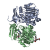
| ||||||||
|---|---|---|---|---|---|---|---|---|---|
| 1 |
| ||||||||
| Unit cell |
|
-Components
-Protein , 2 types, 2 molecules AB
| #1: Protein | Mass: 31451.229 Da / Num. of mol.: 1 Source method: isolated from a genetically manipulated source Source: (gene. exp.) Synechococcus elongatus (strain PCC 7942) (bacteria)Strain: PCC 7942 / Gene: speE, Synpcc7942_0628 / Plasmid: pET22b / Production host: |
|---|---|
| #2: Protein | Mass: 31346.182 Da / Num. of mol.: 1 Source method: isolated from a genetically manipulated source Source: (gene. exp.) Synechococcus elongatus (strain PCC 7942) (bacteria)Strain: PCC 7942 / Gene: speE, Synpcc7942_0628 / Plasmid: pET22b / Production host: |
-Non-polymers , 5 types, 260 molecules 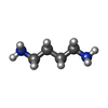


| #3: Chemical | ChemComp-PUT /  Mass: 88.151 Da / Num. of mol.: 1 / Source method: obtained synthetically / Formula: C4H12N2 / Feature type: SUBJECT OF INVESTIGATION Mass: 88.151 Da / Num. of mol.: 1 / Source method: obtained synthetically / Formula: C4H12N2 / Feature type: SUBJECT OF INVESTIGATION |
|---|---|
| #4: Chemical | ChemComp-SPD /  Mass: 145.246 Da / Num. of mol.: 1 / Source method: obtained synthetically / Formula: C7H19N3 / Feature type: SUBJECT OF INVESTIGATION Mass: 145.246 Da / Num. of mol.: 1 / Source method: obtained synthetically / Formula: C7H19N3 / Feature type: SUBJECT OF INVESTIGATION |
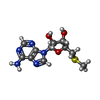
| #5: Chemical | ChemComp-MTA /  Mass: 297.334 Da / Num. of mol.: 1 / Source method: obtained synthetically / Formula: C11H15N5O3S / Feature type: SUBJECT OF INVESTIGATION Mass: 297.334 Da / Num. of mol.: 1 / Source method: obtained synthetically / Formula: C11H15N5O3S / Feature type: SUBJECT OF INVESTIGATION |
| #6: Chemical | ChemComp-PXN / ( Mass: 368.463 Da / Num. of mol.: 1 / Source method: obtained synthetically / Formula: C17H36O8 Mass: 368.463 Da / Num. of mol.: 1 / Source method: obtained synthetically / Formula: C17H36O8 |
| #7: Water | ChemComp-HOH / Mass: 18.015 Da / Num. of mol.: 256 / Source method: isolated from a natural source / Formula: H2O |
-Experimental details
-Experiment
| Experiment | Method: X-RAY DIFFRACTION / Number of used crystals: 1 |
|---|
- Sample preparation
| Crystal | Density Matthews: 2.16 Å3/Da / Density % sol: 43 % |
|---|---|
| Crystal grow | Temperature: 294.15 K / Method: vapor diffusion, sitting drop Details: 0.1 M MES pH 6.0, 45 % v/v Pentaerythritol propoxylate (5/4 PO/OH) |
-Data collection
| Diffraction | Mean temperature: 100 K / Serial crystal experiment: N |
|---|---|
| Diffraction source | Source: SYNCHROTRON / Site: ESRF  / Beamline: MASSIF-3 / Wavelength: 0.9677 Å / Beamline: MASSIF-3 / Wavelength: 0.9677 Å |
| Detector | Type: DECTRIS EIGER X 4M / Detector: PIXEL / Date: Apr 30, 2016 |
| Radiation | Protocol: SINGLE WAVELENGTH / Monochromatic (M) / Laue (L): M / Scattering type: x-ray |
| Radiation wavelength | Wavelength: 0.9677 Å / Relative weight: 1 |
| Reflection | Resolution: 2.18→48.17 Å / Num. obs: 27391 / % possible obs: 99 % / Redundancy: 4.2 % / CC1/2: 0.997 / Rmerge(I) obs: 0.0721 / Rrim(I) all: 0.083 / Net I/σ(I): 16.18 |
| Reflection shell | Resolution: 2.18→2.27 Å / Rmerge(I) obs: 0.226 / CC1/2: 0.956 / Rrim(I) all: 0.261 |
- Processing
| Software |
| |||||||||||||||||||||||||||||||||||||||||||||||||||||||||||||||||||||||||||
|---|---|---|---|---|---|---|---|---|---|---|---|---|---|---|---|---|---|---|---|---|---|---|---|---|---|---|---|---|---|---|---|---|---|---|---|---|---|---|---|---|---|---|---|---|---|---|---|---|---|---|---|---|---|---|---|---|---|---|---|---|---|---|---|---|---|---|---|---|---|---|---|---|---|---|---|---|
| Refinement | Method to determine structure: MOLECULAR REPLACEMENT Starting model: 4YUV Resolution: 2.18→48.17 Å / Cor.coef. Fo:Fc: 0.964 / Cor.coef. Fo:Fc free: 0.939 / Cross valid method: THROUGHOUT / σ(F): 0 / ESU R: 0.256 / ESU R Free: 0.181 Details: HYDROGENS HAVE BEEN ADDED IN THE RIDING POSITIONS U VALUES : REFINED INDIVIDUALLY
| |||||||||||||||||||||||||||||||||||||||||||||||||||||||||||||||||||||||||||
| Solvent computation | Ion probe radii: 0.8 Å / Shrinkage radii: 0.8 Å / VDW probe radii: 1.2 Å | |||||||||||||||||||||||||||||||||||||||||||||||||||||||||||||||||||||||||||
| Displacement parameters | Biso max: 83.54 Å2 / Biso mean: 17.36 Å2 / Biso min: 6.31 Å2
| |||||||||||||||||||||||||||||||||||||||||||||||||||||||||||||||||||||||||||
| Refinement step | Cycle: final / Resolution: 2.18→48.17 Å
| |||||||||||||||||||||||||||||||||||||||||||||||||||||||||||||||||||||||||||
| Refine LS restraints |
| |||||||||||||||||||||||||||||||||||||||||||||||||||||||||||||||||||||||||||
| LS refinement shell | Resolution: 2.181→2.238 Å / Total num. of bins used: 20
|
