 Movie
Movie Controller


+ Open data

- Basic information
| Entry | Database: PDB / ID: 6qk4 | ||||||
|---|---|---|---|---|---|---|---|
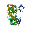
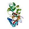
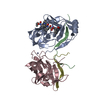
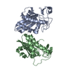
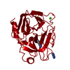
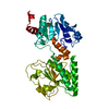
| Title | Lytic transglycosylase, LtgG, of Burkholderia pseudomallei. | ||||||
 Components Components | Membrane-bound lytic murein transglycosylase A | ||||||
 Keywords Keywords | LYASE / lytic transglycosylase / peptidoglycan / cell division / bacterial pathogenesis / Burkholderia pseudomallei | ||||||
| Function / homology |  Function and homology information Function and homology information: / peptidoglycan lytic transglycosylase activity / peptidoglycan turnover / outer membrane / hydrolase activity, hydrolyzing O-glycosyl compounds / peptidoglycan catabolic process / cell wall organization Similarity search - Function | ||||||
| Biological species |  Burkholderia pseudomallei (bacteria) Burkholderia pseudomallei (bacteria) | ||||||
| Method |  X-RAY DIFFRACTION / SYNCHROTRON / MOLECULAR REPLACEMENT / Resolution: 1.73 Å X-RAY DIFFRACTION / SYNCHROTRON / MOLECULAR REPLACEMENT / Resolution: 1.73 Å | ||||||
 Authors Authors | Jenkins, C.H. / Wallis, R. / Allcock, N. / Barnes, K.B. / Richards, M.I. / Auty, J.M. / Galyov, E.E. / Harding, S.V. / Mukamolova, G.V. | ||||||
 Citation Citation | Journal: Sci Rep / Year: 2019 Title: The lytic transglycosylase, LtgG, controls cell morphology and virulence in Burkholderia pseudomallei. Authors: Jenkins, C.H. / Wallis, R. / Allcock, N. / Barnes, K.B. / Richards, M.I. / Auty, J.M. / Galyov, E.E. / Harding, S.V. / Mukamolova, G.V. | ||||||
| History |
|
- Structure visualization
| Structure viewer | Molecule: MolmilJmol/JSmol |
|---|
- Downloads & links
-Download
| PDBx/mmCIF format | 6qk4.cif.gz | 144.4 KB | Display | PDBx/mmCIF format |
|---|---|---|---|---|
| PDB format | pdb6qk4.ent.gz | 111.6 KB | Display | PDB format |
| PDBx/mmJSON format | 6qk4.json.gz | Tree view | PDBx/mmJSON format | |
| Others |  Other downloads Other downloads |
-Validation report
| Arichive directory | https://data.pdbj.org/pub/pdb/validation_reports/qk/6qk4ftp://data.pdbj.org/pub/pdb/validation_reports/qk/6qk4 | HTTPS FTP |
|---|
-Related structure data


-Links
 PDBj
PDBj


- Assembly
| Deposited unit | 
| ||||||||
|---|---|---|---|---|---|---|---|---|---|
| 1 |
| ||||||||
| Unit cell |
|
-Components
| #1: Protein | Mass: 37684.426 Da / Num. of mol.: 1 Source method: isolated from a genetically manipulated source Source: (gene. exp.) Burkholderia pseudomallei (bacteria) / Gene: mltA, CXQ84_03095, DP49_5753, ERS013345_03240 / Production host: References: UniProt: A0A069B8V2, UniProt: Q63QH7*PLUS, Lyases; Carbon-oxygen lyases; Acting on polysaccharides |
|---|---|
| #2: Water | ChemComp-HOH /  Mass: 18.015 Da / Num. of mol.: 237 / Source method: isolated from a natural source / Formula: H2O Mass: 18.015 Da / Num. of mol.: 237 / Source method: isolated from a natural source / Formula: H2O |
| Has protein modification | Y |
-Experimental details
-Experiment
| Experiment | Method: X-RAY DIFFRACTION / Number of used crystals: 1 |
|---|
- Sample preparation
| Crystal | Density Matthews: 2.29 Å3/Da / Density % sol: 46.22 % |
|---|---|
| Crystal grow | Temperature: 277 K / Method: vapor diffusion, sitting drop / pH: 7.5 Details: 0.1 M Bis-tris propane (pH 7.5), containing 0.2 M potassium sodium tartrate and 14% PEG 8K |
-Data collection
| Diffraction | Mean temperature: 100 K / Serial crystal experiment: N |
|---|---|
| Diffraction source | Source: SYNCHROTRON / Site: Diamond  / Beamline: I03 / Wavelength: 0.9763 Å / Beamline: I03 / Wavelength: 0.9763 Å |
| Detector | Type: DECTRIS PILATUS3 S 6M / Detector: PIXEL / Date: Dec 7, 2015 |
| Radiation | Protocol: SINGLE WAVELENGTH / Monochromatic (M) / Laue (L): M / Scattering type: x-ray |
| Radiation wavelength | Wavelength: 0.9763 Å / Relative weight: 1 |
| Reflection | Resolution: 1.73→56.1 Å / Num. obs: 37097 / % possible obs: 99.98 % / Redundancy: 16.3 % / Rsym value: 0.112 / Net I/σ(I): 16.1 |
| Reflection shell | Resolution: 1.73→1.78 Å / Redundancy: 8.6 % / Mean I/σ(I) obs: 2.6 / Rsym value: 0.7 / % possible all: 100 |
- Processing
| Software |
| |||||||||||||||||||||||||||||||||||||||||||||||||||||||||||||||||||||||||||||||||||||||||||||||||||||||||
|---|---|---|---|---|---|---|---|---|---|---|---|---|---|---|---|---|---|---|---|---|---|---|---|---|---|---|---|---|---|---|---|---|---|---|---|---|---|---|---|---|---|---|---|---|---|---|---|---|---|---|---|---|---|---|---|---|---|---|---|---|---|---|---|---|---|---|---|---|---|---|---|---|---|---|---|---|---|---|---|---|---|---|---|---|---|---|---|---|---|---|---|---|---|---|---|---|---|---|---|---|---|---|---|---|---|---|
| Refinement | Method to determine structure: MOLECULAR REPLACEMENT Starting model: 2G5D, 2GAE Resolution: 1.73→56.093 Å / SU ML: 0.16 / Cross valid method: FREE R-VALUE / σ(F): 1.36 / Phase error: 18.99
| |||||||||||||||||||||||||||||||||||||||||||||||||||||||||||||||||||||||||||||||||||||||||||||||||||||||||
| Solvent computation | Shrinkage radii: 0.9 Å / VDW probe radii: 1.11 Å | |||||||||||||||||||||||||||||||||||||||||||||||||||||||||||||||||||||||||||||||||||||||||||||||||||||||||
| Displacement parameters | Biso mean: 34.6 Å2 | |||||||||||||||||||||||||||||||||||||||||||||||||||||||||||||||||||||||||||||||||||||||||||||||||||||||||
| Refinement step | Cycle: LAST / Resolution: 1.73→56.093 Å
| |||||||||||||||||||||||||||||||||||||||||||||||||||||||||||||||||||||||||||||||||||||||||||||||||||||||||
| Refine LS restraints |
| |||||||||||||||||||||||||||||||||||||||||||||||||||||||||||||||||||||||||||||||||||||||||||||||||||||||||
| LS refinement shell |
| |||||||||||||||||||||||||||||||||||||||||||||||||||||||||||||||||||||||||||||||||||||||||||||||||||||||||
| Refinement TLS params. | Method: refined / Origin x: 18.1261 Å / Origin y: -17.5627 Å / Origin z: 14.09 Å
| |||||||||||||||||||||||||||||||||||||||||||||||||||||||||||||||||||||||||||||||||||||||||||||||||||||||||
| Refinement TLS group | Selection details: (chain B and resseq 10:348) |
