 Movie
Movie Controller


+ Open data

- Basic information
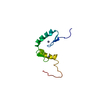
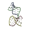
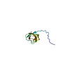
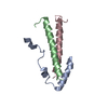
| Entry | Database: PDB / ID: 6qjm | |||||||||
|---|---|---|---|---|---|---|---|---|---|---|
| Title | Cryo-EM structure of heparin-induced 2N4R tau twister filaments | |||||||||
 Components Components | Microtubule-associated protein tau | |||||||||
 Keywords Keywords | PROTEIN FIBRIL / Recombinant tau protein / heparin / filament / cross-beta structure | |||||||||
| Function / homology |  Function and homology information Function and homology informationplus-end-directed organelle transport along microtubule / histone-dependent DNA binding / negative regulation of protein localization to mitochondrion / neurofibrillary tangle / microtubule lateral binding / axonal transport / tubulin complex / positive regulation of protein localization to synapse / phosphatidylinositol bisphosphate binding / generation of neurons ...plus-end-directed organelle transport along microtubule / histone-dependent DNA binding / negative regulation of protein localization to mitochondrion / neurofibrillary tangle / microtubule lateral binding / axonal transport / tubulin complex / positive regulation of protein localization to synapse / phosphatidylinositol bisphosphate binding / generation of neurons / rRNA metabolic process / axonal transport of mitochondrion / regulation of mitochondrial fission / axon development / regulation of microtubule-based movement / intracellular distribution of mitochondria / regulation of chromosome organization / central nervous system neuron development / minor groove of adenine-thymine-rich DNA binding / lipoprotein particle binding / microtubule polymerization / negative regulation of mitochondrial membrane potential / regulation of microtubule polymerization / dynactin binding / apolipoprotein binding / protein polymerization / main axon / Caspase-mediated cleavage of cytoskeletal proteins / regulation of microtubule polymerization or depolymerization / negative regulation of mitochondrial fission / axolemma / glial cell projection / neurofibrillary tangle assembly / positive regulation of axon extension / regulation of cellular response to heat / positive regulation of microtubule polymerization / positive regulation of protein localization / Activation of AMPK downstream of NMDARs / positive regulation of superoxide anion generation / cellular response to brain-derived neurotrophic factor stimulus / regulation of long-term synaptic depression / supramolecular fiber organization / cytoplasmic microtubule organization / regulation of calcium-mediated signaling / axon cytoplasm / somatodendritic compartment / synapse assembly / phosphatidylinositol binding / nuclear periphery / astrocyte activation / enzyme inhibitor activity / protein phosphatase 2A binding / stress granule assembly / regulation of microtubule cytoskeleton organization / regulation of autophagy / cellular response to reactive oxygen species / microglial cell activation / cellular response to nerve growth factor stimulus / Hsp90 protein binding / protein homooligomerization / SH3 domain binding / PKR-mediated signaling / synapse organization / regulation of synaptic plasticity / response to lead ion / microtubule cytoskeleton organization / memory / neuron projection development / cytoplasmic ribonucleoprotein granule / cell-cell signaling / single-stranded DNA binding / cellular response to heat / growth cone / protein-folding chaperone binding / microtubule cytoskeleton / actin binding / cell body / double-stranded DNA binding / sequence-specific DNA binding / microtubule binding / amyloid fibril formation / dendritic spine / microtubule / protein-macromolecule adaptor activity / learning or memory / neuron projection / membrane raft / negative regulation of gene expression / axon / neuronal cell body / DNA damage response / dendrite / protein kinase binding / enzyme binding / mitochondrion / DNA binding / RNA binding / extracellular region / identical protein binding / nucleus Similarity search - Function | |||||||||
| Biological species |  Homo sapiens (human) Homo sapiens (human) | |||||||||
| Method | ELECTRON MICROSCOPY / single particle reconstruction / cryo EM / Resolution: 3.3 Å | |||||||||
 Authors Authors | Zhang, W. / Falcon, B. / Murzin, A.G. / Fan, J. / Crowther, R.A. / Goedert, M. / Scheres, S.H.W. | |||||||||
| Funding support |  United Kingdom, 2items United Kingdom, 2items
| |||||||||
 Citation Citation | Journal: Elife / Year: 2019 Title: Heparin-induced tau filaments are polymorphic and differ from those in Alzheimer's and Pick's diseases. Authors: Wenjuan Zhang / Benjamin Falcon / Alexey G Murzin / Juan Fan / R Anthony Crowther / Michel Goedert / Sjors Hw Scheres / Abstract: Assembly of microtubule-associated protein tau into filamentous inclusions underlies a range of neurodegenerative diseases. Tau filaments adopt different conformations in Alzheimer's and Pick's ...Assembly of microtubule-associated protein tau into filamentous inclusions underlies a range of neurodegenerative diseases. Tau filaments adopt different conformations in Alzheimer's and Pick's diseases. Here, we used cryo- and immuno- electron microscopy to characterise filaments that were assembled from recombinant full-length human tau with four (2N4R) or three (2N3R) microtubule-binding repeats in the presence of heparin. 2N4R tau assembles into multiple types of filaments, and the structures of three types reveal similar 'kinked hairpin' folds, in which the second and third repeats pack against each other. 2N3R tau filaments are structurally homogeneous, and adopt a dimeric core, where the third repeats of two tau molecules pack in a parallel manner. The heparin-induced tau filaments differ from those of Alzheimer's or Pick's disease, which have larger cores with different repeat compositions. Our results illustrate the structural versatility of amyloid filaments, and raise questions about the relevance of in vitro assembly. | |||||||||
| History |
|
- Structure visualization
| Movie |
Movie viewer |
|---|---|
| Structure viewer | Molecule: MolmilJmol/JSmol |
- Downloads & links
-Download
| PDBx/mmCIF format | 6qjm.cif.gz | 30.4 KB | Display | PDBx/mmCIF format |
|---|---|---|---|---|
| PDB format | pdb6qjm.ent.gz | 19.9 KB | Display | PDB format |
| PDBx/mmJSON format | 6qjm.json.gz | Tree view | PDBx/mmJSON format | |
| Others |  Other downloads Other downloads |
-Validation report
| Arichive directory | https://data.pdbj.org/pub/pdb/validation_reports/qj/6qjmftp://data.pdbj.org/pub/pdb/validation_reports/qj/6qjm | HTTPS FTP |
|---|
-Related structure data
| Related structure data | 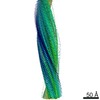 4564MC 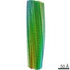 4563C 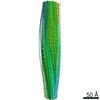 4565C 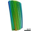 4566C 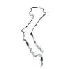 6qjhC  6qjpC  6qjqC M: map data used to model this data C: citing same article ( |
|---|---|
| Similar structure data | |
| EM raw data | EMPIAR-10243 (Title: Cryo-EM reconstruction of heparin-induced 2N4R tau filaments Data size: 446.3 / Data #1: Aligned micrographs [micrographs - single frame] / Data #2: Raw movies [micrographs - multiframe]) |


-Links
 PDBj
PDBj


- Assembly
| Deposited unit | 
|
|---|---|
| 1 |
|
-Components
| #1: Protein/peptide | Mass: 5189.081 Da / Num. of mol.: 3 Source method: isolated from a genetically manipulated source Source: (gene. exp.) Homo sapiens (human) / Gene: MAPT, MAPTL, MTBT1, TAU / Plasmid: pRK172 / Production host:  |
|---|
-Experimental details
-Experiment
| Experiment | Method: ELECTRON MICROSCOPY |
|---|---|
| EM experiment | Aggregation state: FILAMENT / 3D reconstruction method: single particle reconstruction |
- Sample preparation
| Component | Name: heparin-induced 2N4R tau twister filaments / Type: COMPLEX Details: Recombinant tau protein was induced into filaments by incubation with heparin at 37 C for 3 days Entity ID: all / Source: RECOMBINANT | |||||||||||||||
|---|---|---|---|---|---|---|---|---|---|---|---|---|---|---|---|---|
| Molecular weight | Value: 45.85 kDa/nm / Experimental value: NO | |||||||||||||||
| Source (natural) | Organism: Homo sapiens (human) | |||||||||||||||
| Source (recombinant) | Organism: | |||||||||||||||
| Buffer solution | pH: 7.4 / Details: 20 mM Tris, pH 7.4, 100mM NaCl | |||||||||||||||
| Buffer component |
| |||||||||||||||
| Specimen | Conc.: 2 mg/ml / Embedding applied: NO / Shadowing applied: NO / Staining applied: NO / Vitrification applied: YES Details: Recombinant tau protein was induced into filaments by incubation with heparin at 37 C for 3 days. The filaments were pronase-treated before making Cryo-grids. | |||||||||||||||
| Specimen support | Grid material: GOLD / Grid mesh size: 300 divisions/in. / Grid type: Quantifoil R1.2/1.3 | |||||||||||||||
| Vitrification | Instrument: FEI VITROBOT MARK IV / Cryogen name: ETHANE / Humidity: 100 % / Chamber temperature: 277 K / Details: Blot force: -12 ; Blot time: 4s |
- Electron microscopy imaging
| Experimental equipment |  Model: Tecnai Polara / Image courtesy: FEI Company |
|---|---|
| Microscopy | Model: FEI POLARA 300 |
| Electron gun | Electron source:  FIELD EMISSION GUN / Accelerating voltage: 300 kV / Illumination mode: FLOOD BEAM FIELD EMISSION GUN / Accelerating voltage: 300 kV / Illumination mode: FLOOD BEAM |
| Electron lens | Mode: BRIGHT FIELD / Nominal defocus max: 2800 nm / Nominal defocus min: 1700 nm / Cs: 2 mm / C2 aperture diameter: 50 µm / Alignment procedure: COMA FREE |
| Specimen holder | Cryogen: NITROGEN / Specimen holder model: GATAN LIQUID NITROGEN |
| Image recording | Average exposure time: 1 sec. / Electron dose: 48 e/Å2 / Detector mode: INTEGRATING / Film or detector model: FEI FALCON III (4k x 4k) / Num. of grids imaged: 1 / Num. of real images: 717 Details: Images were collected in movie-mode at 30 frames per second |
| Image scans | Sampling size: 14 µm / Width: 4096 / Height: 4096 |
- Processing
| EM software |
| ||||||||||||||||||||||||||||||||||||||||||||
|---|---|---|---|---|---|---|---|---|---|---|---|---|---|---|---|---|---|---|---|---|---|---|---|---|---|---|---|---|---|---|---|---|---|---|---|---|---|---|---|---|---|---|---|---|---|
| Image processing | Details: Movie frames were gain-corrected, aligned, dose weighted and then summed into a single micrograph using MOTIONCOR2 (Zheng et al., 2017) | ||||||||||||||||||||||||||||||||||||||||||||
| CTF correction | Details: Aligned, non-dose-weighted micrographs were used to estimate the contrast transfer function (CTF) using CTFFIND4.1 Type: PHASE FLIPPING AND AMPLITUDE CORRECTION | ||||||||||||||||||||||||||||||||||||||||||||
| Helical symmerty | Angular rotation/subunit: -3.38 ° / Axial rise/subunit: 4.7 Å / Axial symmetry: C1 | ||||||||||||||||||||||||||||||||||||||||||||
| Particle selection | Num. of particles selected: 187555 / Details: Manually picked | ||||||||||||||||||||||||||||||||||||||||||||
| 3D reconstruction | Resolution: 3.3 Å / Resolution method: FSC 0.143 CUT-OFF / Num. of particles: 141461 / Algorithm: FOURIER SPACE / Details: we performed two rounds of 3D auto-refinement. / Num. of class averages: 6 / Symmetry type: HELICAL | ||||||||||||||||||||||||||||||||||||||||||||
| Atomic model building | B value: 58.51 / Protocol: AB INITIO MODEL / Space: RECIPROCAL / Target criteria: Fourier shell correlation Details: A stack of three consecutive monomers was refined to preserve nearest-neighbour interactions for the middle chain. Side-chain clashes were detected using MOLPROBITY, and corrected by ...Details: A stack of three consecutive monomers was refined to preserve nearest-neighbour interactions for the middle chain. Side-chain clashes were detected using MOLPROBITY, and corrected by iterative cycles of real-space refinement in COOT and Fourier-space refinement in REFMAC and PHENIX. For each refined structure, separate model refinements were performed against a single half-map, and the resulting model was compared to the other half-map to confirm the absence of overfitting. |
