 Movie
Movie Controller

[English] 日本語
 Yorodumi
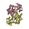
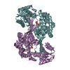
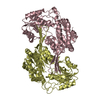
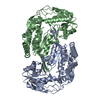
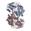
- PDB-6qhn: Metagenome-derived salicylaldehyde dehydrogenase from alpine soil... -
+ Open data

- Basic information
| Entry | Database: PDB / ID: 6qhn | ||||||
|---|---|---|---|---|---|---|---|
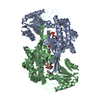
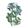
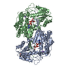
| Title | Metagenome-derived salicylaldehyde dehydrogenase from alpine soil in complex with protocatechuic acid | ||||||
 Components Components | SALICYLALDEHYDE DEHYDROGENASE | ||||||
 Keywords Keywords | OXIDOREDUCTASE / salicylaldehyde dehydrogenase / metagenome / alpine soil / complex | ||||||
| Function / homology | Aldehyde Dehydrogenase; Chain A, domain 1 / Aldehyde Dehydrogenase; Chain A, domain 1 / 3-Layer(aba) Sandwich / Alpha Beta / 3,4-DIHYDROXYBENZOIC ACID Function and homology information Function and homology information | ||||||
| Biological species |  Alphaproteobacteria (a-proteobacteria) Alphaproteobacteria (a-proteobacteria) | ||||||
| Method |  X-RAY DIFFRACTION / SYNCHROTRON / MOLECULAR REPLACEMENT / molecular replacement / Resolution: 1.9 Å X-RAY DIFFRACTION / SYNCHROTRON / MOLECULAR REPLACEMENT / molecular replacement / Resolution: 1.9 Å | ||||||
 Authors Authors | Hakansson, M. / Svensson, L.A. / Shamsudeen, D.U. / Allen, C.C.R. | ||||||
 Citation Citation | Journal: Acta Crystallogr.,Sect.F / Year: 2022 Title: Expression, purification and crystallization of a novel metagenome-derived salicylaldehyde dehydrogenase from Alpine soil. Authors: Dandare, S.U. / Hakansson, M. / Svensson, L.A. / Timson, D.J. / Allen, C.C.R. | ||||||
| History |
|
- Structure visualization
| Structure viewer | Molecule: MolmilJmol/JSmol |
|---|
- Downloads & links
-Download
| PDBx/mmCIF format | 6qhn.cif.gz | 717.2 KB | Display | PDBx/mmCIF format |
|---|---|---|---|---|
| PDB format | pdb6qhn.ent.gz | 595.7 KB | Display | PDB format |
| PDBx/mmJSON format | 6qhn.json.gz | Tree view | PDBx/mmJSON format | |
| Others |  Other downloads Other downloads |
-Validation report
| Arichive directory | https://data.pdbj.org/pub/pdb/validation_reports/qh/6qhnftp://data.pdbj.org/pub/pdb/validation_reports/qh/6qhn | HTTPS FTP |
|---|
-Related structure data
| Related structure data | 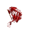 4jz6S S: Starting model for refinement |
|---|---|
| Similar structure data |


-Links
 PDBj
PDBj- Assembly
| Deposited unit | 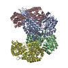
| |||||||||
|---|---|---|---|---|---|---|---|---|---|---|
| 1 | 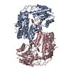
| |||||||||
| 2 | 
| |||||||||
| Unit cell |
| |||||||||
| Components on special symmetry positions |
|
-Components
| #1: Protein | Mass: 49128.480 Da / Num. of mol.: 4 Source method: isolated from a genetically manipulated source Source: (gene. exp.) Alphaproteobacteria (a-proteobacteria) / Production host: #2: Chemical | ChemComp-DHB / 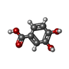  Mass: 154.120 Da / Num. of mol.: 4 / Source method: obtained synthetically / Formula: C7H6O4 / Feature type: SUBJECT OF INVESTIGATION Mass: 154.120 Da / Num. of mol.: 4 / Source method: obtained synthetically / Formula: C7H6O4 / Feature type: SUBJECT OF INVESTIGATION#3: Chemical | ChemComp-GOL / |   Mass: 92.094 Da / Num. of mol.: 1 / Source method: obtained synthetically / Formula: C3H8O3 Mass: 92.094 Da / Num. of mol.: 1 / Source method: obtained synthetically / Formula: C3H8O3#4: Water | ChemComp-HOH / |  Mass: 18.015 Da / Num. of mol.: 1609 / Source method: isolated from a natural source / Formula: H2O Mass: 18.015 Da / Num. of mol.: 1609 / Source method: isolated from a natural source / Formula: H2OHas ligand of interest | Y | |
|---|
-Experimental details
-Experiment
| Experiment | Method: X-RAY DIFFRACTION / Number of used crystals: 1 |
|---|
- Sample preparation
| Crystal | Density Matthews: 2.88 Å3/Da / Density % sol: 57.25 % |
|---|---|
| Crystal grow | Temperature: 293 K / Method: vapor diffusion, sitting drop / pH: 4.6 / Details: 0.1 M sodium acetate pH 4.6 and 8 % w/v PEG 8000 |
-Data collection
| Diffraction | Mean temperature: 100 K / Serial crystal experiment: N | |||||||||||||||||||||||||||
|---|---|---|---|---|---|---|---|---|---|---|---|---|---|---|---|---|---|---|---|---|---|---|---|---|---|---|---|---|
| Diffraction source | Source: SYNCHROTRON / Site: Diamond  / Beamline: I03 / Wavelength: 0.97629 Å / Beamline: I03 / Wavelength: 0.97629 Å | |||||||||||||||||||||||||||
| Detector | Type: DECTRIS PILATUS3 6M / Detector: PIXEL / Date: Oct 5, 2017 | |||||||||||||||||||||||||||
| Radiation | Protocol: SINGLE WAVELENGTH / Monochromatic (M) / Laue (L): M / Scattering type: x-ray | |||||||||||||||||||||||||||
| Radiation wavelength | Wavelength: 0.97629 Å / Relative weight: 1 | |||||||||||||||||||||||||||
| Reflection | Resolution: 1.9→29.89 Å / Num. obs: 177084 / % possible obs: 99.9 % / Redundancy: 5.6 % / Biso Wilson estimate: 31.44 Å2 / CC1/2: 0.997 / Rmerge(I) obs: 0.13 / Rpim(I) all: 0.061 / Rrim(I) all: 0.144 / Net I/σ(I): 8.7 | |||||||||||||||||||||||||||
| Reflection shell | Diffraction-ID: 1
|
-Phasing
| Phasing | Method: molecular replacement | |||||||||
|---|---|---|---|---|---|---|---|---|---|---|
| Phasing MR | Model details: Phaser MODE: MR_AUTO
|
- Processing
| Software |
| |||||||||||||||||||||||||||||||||||||||||||||||||||||||||||||||||||||||||||||||||||||||||||||||||||||||||||||||||||||||||||||
|---|---|---|---|---|---|---|---|---|---|---|---|---|---|---|---|---|---|---|---|---|---|---|---|---|---|---|---|---|---|---|---|---|---|---|---|---|---|---|---|---|---|---|---|---|---|---|---|---|---|---|---|---|---|---|---|---|---|---|---|---|---|---|---|---|---|---|---|---|---|---|---|---|---|---|---|---|---|---|---|---|---|---|---|---|---|---|---|---|---|---|---|---|---|---|---|---|---|---|---|---|---|---|---|---|---|---|---|---|---|---|---|---|---|---|---|---|---|---|---|---|---|---|---|---|---|---|
| Refinement | Method to determine structure: MOLECULAR REPLACEMENT Starting model: 4JZ6 Resolution: 1.9→27.67 Å / Cor.coef. Fo:Fc: 0.959 / Cor.coef. Fo:Fc free: 0.948 / SU R Cruickshank DPI: 0.174 / Cross valid method: THROUGHOUT / σ(F): 0 / SU R Blow DPI: 0.132 / SU Rfree Blow DPI: 0.118 / SU Rfree Cruickshank DPI: 0.115
| |||||||||||||||||||||||||||||||||||||||||||||||||||||||||||||||||||||||||||||||||||||||||||||||||||||||||||||||||||||||||||||
| Displacement parameters | Biso max: 183.02 Å2 / Biso mean: 37.33 Å2 / Biso min: 5.83 Å2
| |||||||||||||||||||||||||||||||||||||||||||||||||||||||||||||||||||||||||||||||||||||||||||||||||||||||||||||||||||||||||||||
| Refine analyze | Luzzati coordinate error obs: 0.23 Å | |||||||||||||||||||||||||||||||||||||||||||||||||||||||||||||||||||||||||||||||||||||||||||||||||||||||||||||||||||||||||||||
| Refinement step | Cycle: final / Resolution: 1.9→27.67 Å
| |||||||||||||||||||||||||||||||||||||||||||||||||||||||||||||||||||||||||||||||||||||||||||||||||||||||||||||||||||||||||||||
| Refine LS restraints |
| |||||||||||||||||||||||||||||||||||||||||||||||||||||||||||||||||||||||||||||||||||||||||||||||||||||||||||||||||||||||||||||
| LS refinement shell | Resolution: 1.9→1.91 Å / Rfactor Rfree error: 0 / Total num. of bins used: 50
| |||||||||||||||||||||||||||||||||||||||||||||||||||||||||||||||||||||||||||||||||||||||||||||||||||||||||||||||||||||||||||||
| Refinement TLS params. | Method: refined / Refine-ID: X-RAY DIFFRACTION
| |||||||||||||||||||||||||||||||||||||||||||||||||||||||||||||||||||||||||||||||||||||||||||||||||||||||||||||||||||||||||||||
| Refinement TLS group |
|
