 Movie
Movie Controller


+ Open data

- Basic information
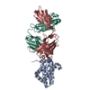
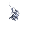
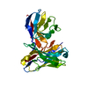
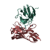
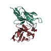
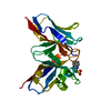
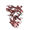
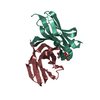
| Entry | Database: PDB / ID: 6qf6 | ||||||
|---|---|---|---|---|---|---|---|
| Title | Structure of an E.coli expressed anti-Mcl1 scFv | ||||||
 Components Components | E.coli expressed scFv | ||||||
 Keywords Keywords | APOPTOSIS / Mcl1 / scFv / AZD5991 | ||||||
| Function / homology | Immunoglobulins / Immunoglobulin-like / Sandwich / Mainly Beta Function and homology information Function and homology information | ||||||
| Biological species |  Homo sapiens (human) Homo sapiens (human) | ||||||
| Method |  X-RAY DIFFRACTION / SYNCHROTRON / MOLECULAR REPLACEMENT / Resolution: 2.59 Å X-RAY DIFFRACTION / SYNCHROTRON / MOLECULAR REPLACEMENT / Resolution: 2.59 Å | ||||||
 Authors Authors | Luptak, J. | ||||||
 Citation Citation | Journal: To Be Published Title: Structure of an E.coli expressed anti-Mcl1 scFv Authors: Luptak, J. | ||||||
| History |
|
- Structure visualization
| Structure viewer | Molecule: MolmilJmol/JSmol |
|---|
- Downloads & links
-Download
| PDBx/mmCIF format | 6qf6.cif.gz | 181.5 KB | Display | PDBx/mmCIF format |
|---|---|---|---|---|
| PDB format | pdb6qf6.ent.gz | 145.4 KB | Display | PDB format |
| PDBx/mmJSON format | 6qf6.json.gz | Tree view | PDBx/mmJSON format | |
| Others |  Other downloads Other downloads |
-Validation report
| Summary document | 6qf6_validation.pdf.gz | 450.1 KB | Display | wwPDB validaton report |
|---|---|---|---|---|
| Full document | 6qf6_full_validation.pdf.gz | 462.8 KB | Display | |
| Data in XML | 6qf6_validation.xml.gz | 33.8 KB | Display | |
| Data in CIF | 6qf6_validation.cif.gz | 48.1 KB | Display | |
| Arichive directory | https://data.pdbj.org/pub/pdb/validation_reports/qf/6qf6ftp://data.pdbj.org/pub/pdb/validation_reports/qf/6qf6 | HTTPS FTP |
-Related structure data
| Related structure data |  6qb3S S: Starting model for refinement |
|---|---|
| Similar structure data |


-Links
 PDBj
PDBj

- Assembly
| Deposited unit | 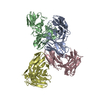
| ||||||||
|---|---|---|---|---|---|---|---|---|---|
| 1 | 
| ||||||||
| 2 | 
| ||||||||
| 3 | 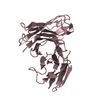
| ||||||||
| 4 | 
| ||||||||
| Unit cell |
|
-Components
| #1: Antibody | Mass: 25507.953 Da / Num. of mol.: 4 Source method: isolated from a genetically manipulated source Source: (gene. exp.) Homo sapiens (human) / Production host:  #2: Water | ChemComp-HOH / |  Mass: 18.015 Da / Num. of mol.: 231 / Source method: isolated from a natural source / Formula: H2O Mass: 18.015 Da / Num. of mol.: 231 / Source method: isolated from a natural source / Formula: H2OHas protein modification | Y | |
|---|
-Experimental details
-Experiment
| Experiment | Method: X-RAY DIFFRACTION / Number of used crystals: 1 |
|---|
- Sample preparation
| Crystal | Density Matthews: 3.71 Å3/Da / Density % sol: 66.85 % |
|---|---|
| Crystal grow | Temperature: 293 K / Method: vapor diffusion, sitting drop Details: 0.4 M Ammonium Sulphate, 25% PEG 3350, 0.1 M BIS-TRIS pH 5.9 |
-Data collection
| Diffraction | Mean temperature: 100 K / Serial crystal experiment: N |
|---|---|
| Diffraction source | Source: SYNCHROTRON / Site: Diamond  / Beamline: I03 / Wavelength: 0.9763 Å / Beamline: I03 / Wavelength: 0.9763 Å |
| Detector | Type: DECTRIS PILATUS3 S 6M / Detector: PIXEL / Date: Nov 24, 2017 |
| Radiation | Protocol: SINGLE WAVELENGTH / Monochromatic (M) / Laue (L): M / Scattering type: x-ray |
| Radiation wavelength | Wavelength: 0.9763 Å / Relative weight: 1 |
| Reflection | Resolution: 2.59→127.403 Å / Num. obs: 23417 / % possible obs: 91.27 % / Redundancy: 12.92 % / Biso Wilson estimate: 42.46 Å2 / CC1/2: 0.978 / Net I/σ(I): 5.75 |
| Reflection shell | Resolution: 2.59→3.01 Å / Num. unique obs: 26817 / CC1/2: 0.369 |
- Processing
| Software |
| ||||||||||||||||||||
|---|---|---|---|---|---|---|---|---|---|---|---|---|---|---|---|---|---|---|---|---|---|
| Refinement | Method to determine structure: MOLECULAR REPLACEMENT Starting model: 6qb3 Resolution: 2.59→127.4 Å / Cross valid method: THROUGHOUT
| ||||||||||||||||||||
| Displacement parameters | Biso max: 119.01 Å2 / Biso mean: 35.9746 Å2 / Biso min: 3 Å2 | ||||||||||||||||||||
| Refinement step | Cycle: LAST / Resolution: 2.59→127.4 Å
|
