 Movie
Movie Controller

[English] 日本語
 Yorodumi
- PDB-6q30: Crystal structure of NDM-1 beta-lactamase in complex with boronic... -
+ Open data

- Basic information
| Entry | Database: PDB / ID: 6q30 | ||||||
|---|---|---|---|---|---|---|---|
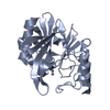
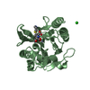
| Title | Crystal structure of NDM-1 beta-lactamase in complex with boronic inhibitor cpd 5 | ||||||
 Components Components | Metallo-beta-lactamase type 2 | ||||||
 Keywords Keywords | HYDROLASE / beta-lactamase / bacterial resistance / acyclic boronic inhibitors | ||||||
| Function / homology |  Function and homology information Function and homology informationantibiotic catabolic process / beta-lactamase activity / beta-lactamase / periplasmic space / response to antibiotic / zinc ion binding Similarity search - Function | ||||||
| Biological species |  Klebsiella pneumoniae (bacteria) Klebsiella pneumoniae (bacteria) | ||||||
| Method |  X-RAY DIFFRACTION / SYNCHROTRON / MOLECULAR REPLACEMENT / Resolution: 1.5 Å X-RAY DIFFRACTION / SYNCHROTRON / MOLECULAR REPLACEMENT / Resolution: 1.5 Å | ||||||
 Authors Authors | Maso, L. / Quotadamo, A. / Bellio, P. / Montanari, M. / Celenza, G. / Venturelli, A. / Costi, M.P. / Tondi, D. / Cendron, L. | ||||||
| Funding support |  Italy, 1items Italy, 1items
| ||||||
 Citation Citation | Journal: Acs Med.Chem.Lett. / Year: 2019 Title: X-ray Crystallography Deciphers the Activity of Broad-Spectrum Boronic Acid beta-Lactamase Inhibitors. Authors: Cendron, L. / Quotadamo, A. / Maso, L. / Bellio, P. / Montanari, M. / Celenza, G. / Venturelli, A. / Costi, M.P. / Tondi, D. | ||||||
| History |
|
- Structure visualization
| Structure viewer | Molecule: MolmilJmol/JSmol |
|---|
- Downloads & links
-Download
| PDBx/mmCIF format | 6q30.cif.gz | 207 KB | Display | PDBx/mmCIF format |
|---|---|---|---|---|
| PDB format | pdb6q30.ent.gz | 162.7 KB | Display | PDB format |
| PDBx/mmJSON format | 6q30.json.gz | Tree view | PDBx/mmJSON format | |
| Others |  Other downloads Other downloads |
-Validation report
| Arichive directory | https://data.pdbj.org/pub/pdb/validation_reports/q3/6q30ftp://data.pdbj.org/pub/pdb/validation_reports/q3/6q30 | HTTPS FTP |
|---|
-Related structure data
| Related structure data |  6ibsC  6ibvC  6q2yC  6q35C  5gzuS S: Starting model for refinement C: citing same article ( |
|---|---|
| Similar structure data |


-Links
 PDBj
PDBj

- Assembly
| Deposited unit | 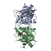
| ||||||||
|---|---|---|---|---|---|---|---|---|---|
| 1 | 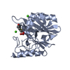
| ||||||||
| 2 | 
| ||||||||
| Unit cell |
|
-Components
| #1: Protein | Mass: 25860.133 Da / Num. of mol.: 2 Source method: isolated from a genetically manipulated source Source: (gene. exp.) Klebsiella pneumoniae (bacteria) / Gene: blaNDM-1 / Production host: #2: Chemical | ChemComp-ZN /   Mass: 65.409 Da / Num. of mol.: 4 / Source method: obtained synthetically / Formula: Zn Mass: 65.409 Da / Num. of mol.: 4 / Source method: obtained synthetically / Formula: Zn#3: Chemical | ChemComp-CA /   Mass: 40.078 Da / Num. of mol.: 4 / Source method: obtained synthetically / Formula: Ca Mass: 40.078 Da / Num. of mol.: 4 / Source method: obtained synthetically / Formula: Ca#4: Chemical |   Mass: 239.033 Da / Num. of mol.: 2 / Source method: obtained synthetically / Formula: C9H8BO5S Mass: 239.033 Da / Num. of mol.: 2 / Source method: obtained synthetically / Formula: C9H8BO5S#5: Water | ChemComp-HOH / |  Mass: 18.015 Da / Num. of mol.: 368 / Source method: isolated from a natural source / Formula: H2O Mass: 18.015 Da / Num. of mol.: 368 / Source method: isolated from a natural source / Formula: H2O |
|---|
-Experimental details
-Experiment
| Experiment | Method: X-RAY DIFFRACTION / Number of used crystals: 1 |
|---|
- Sample preparation
| Crystal | Density Matthews: 2.08 Å3/Da / Density % sol: 40.8 % |
|---|---|
| Crystal grow | Temperature: 293 K / Method: vapor diffusion, sitting drop / pH: 6.3 Details: 0.3 M MgCl2 exahydrate; 0.3 M CaCl2 dihydrate; 25% w/v PEG 3350; 25% w/v MPD; 20% v/v PEG 1000; 0.1 M MES/IMIDAZOLE pH 6.5 |
-Data collection
| Diffraction | Mean temperature: 100 K / Serial crystal experiment: N |
|---|---|
| Diffraction source | Source: SYNCHROTRON / Site: ESRF  / Beamline: ID23-1 / Wavelength: 1 Å / Beamline: ID23-1 / Wavelength: 1 Å |
| Detector | Type: DECTRIS PILATUS3 6M / Detector: PIXEL / Date: Jul 22, 2018 |
| Radiation | Protocol: SINGLE WAVELENGTH / Monochromatic (M) / Laue (L): M / Scattering type: x-ray |
| Radiation wavelength | Wavelength: 1 Å / Relative weight: 1 |
| Reflection | Resolution: 1.5→42.52 Å / Num. obs: 64514 / % possible obs: 98 % / Redundancy: 4.5 % / Biso Wilson estimate: 11.16 Å2 / CC1/2: 0.999 / Rmerge(I) obs: 0.039 / Net I/σ(I): 20.3 |
| Reflection shell | Resolution: 1.5→1.554 Å / Redundancy: 4.5 % / Rmerge(I) obs: 0.426 / Mean I/σ(I) obs: 3.48 / Num. unique obs: 6250 / CC1/2: 0.836 / % possible all: 97.48 |
- Processing
| Software |
| ||||||||||||||||||||||||||||||||||||||||||||||||||||||||||||||||||||||||||||||||||||||||||||||||||||||||||||||||||||||||||||||||||||||||||||||||||||||||||||||||||||||||||||||||||||||
|---|---|---|---|---|---|---|---|---|---|---|---|---|---|---|---|---|---|---|---|---|---|---|---|---|---|---|---|---|---|---|---|---|---|---|---|---|---|---|---|---|---|---|---|---|---|---|---|---|---|---|---|---|---|---|---|---|---|---|---|---|---|---|---|---|---|---|---|---|---|---|---|---|---|---|---|---|---|---|---|---|---|---|---|---|---|---|---|---|---|---|---|---|---|---|---|---|---|---|---|---|---|---|---|---|---|---|---|---|---|---|---|---|---|---|---|---|---|---|---|---|---|---|---|---|---|---|---|---|---|---|---|---|---|---|---|---|---|---|---|---|---|---|---|---|---|---|---|---|---|---|---|---|---|---|---|---|---|---|---|---|---|---|---|---|---|---|---|---|---|---|---|---|---|---|---|---|---|---|---|---|---|---|---|
| Refinement | Method to determine structure: MOLECULAR REPLACEMENT Starting model: 5GZU Resolution: 1.5→42.52 Å / Cor.coef. Fo:Fc: 0.972 / Cor.coef. Fo:Fc free: 0.951 / SU B: 3.787 / SU ML: 0.061 / Cross valid method: THROUGHOUT / ESU R: 0.085 / ESU R Free: 0.075 / Stereochemistry target values: MAXIMUM LIKELIHOOD
| ||||||||||||||||||||||||||||||||||||||||||||||||||||||||||||||||||||||||||||||||||||||||||||||||||||||||||||||||||||||||||||||||||||||||||||||||||||||||||||||||||||||||||||||||||||||
| Solvent computation | Ion probe radii: 0.8 Å / Shrinkage radii: 0.8 Å / VDW probe radii: 1.2 Å / Solvent model: MASK | ||||||||||||||||||||||||||||||||||||||||||||||||||||||||||||||||||||||||||||||||||||||||||||||||||||||||||||||||||||||||||||||||||||||||||||||||||||||||||||||||||||||||||||||||||||||
| Displacement parameters | Biso mean: 18.388 Å2
| ||||||||||||||||||||||||||||||||||||||||||||||||||||||||||||||||||||||||||||||||||||||||||||||||||||||||||||||||||||||||||||||||||||||||||||||||||||||||||||||||||||||||||||||||||||||
| Refinement step | Cycle: LAST / Resolution: 1.5→42.52 Å
| ||||||||||||||||||||||||||||||||||||||||||||||||||||||||||||||||||||||||||||||||||||||||||||||||||||||||||||||||||||||||||||||||||||||||||||||||||||||||||||||||||||||||||||||||||||||
| Refine LS restraints |
|