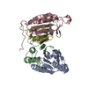
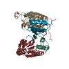
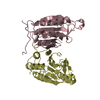
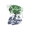
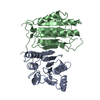
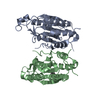
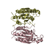
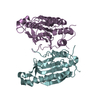
Entry Database : PDB / ID : 6px9Title Crystal structure of procaspase-8 in complex with covalent small molecule inhibitor 63-R Caspase-8 Keywords / / / / Function / homology Function Domain/homology Component
/ / / / / / / / / / / / / / / / / / / / / / / / / / / / / / / / / / / / / / / / / / / / / / / / / / / / / / / / / / / / / / / / / / / / / / / / / / / / / / / / / / / / / / / / / / / / / / / / / / / / / / / / / / / / / / / / / / / / / / / / / / / Biological species Homo sapiens (human)Method / / / Resolution : 2.88 Å Authors Xu, J.H. / Wolan, D.W. Funding support Organization Grant number Country National Institutes of Health/National Institute of General Medical Sciences (NIH/NIGMS) R01GM118382 National Institutes of Health/National Institute of General Medical Sciences (NIH/NIGMS) R01GM069832 Department of Energy (DOE, United States) DE-FC02-02ER63421
Journal : Acs Chem.Biol. / Year : 2020Title : Integrative X-ray Structure and Molecular Modeling for the Rationalization of Procaspase-8 Inhibitor Potency and Selectivity.Authors : Xu, J.H. / Eberhardt, J. / Hill-Payne, B. / Gonzalez-Paez, G.E. / Castellon, J.O. / Cravatt, B.F. / Forli, S. / Wolan, D.W. / Backus, K.M. History Deposition Jul 25, 2019 Deposition site / Processing site Revision 1.0 Jan 29, 2020 Provider / Type Revision 1.1 Mar 4, 2020 Group / Category Item _citation.journal_volume / _citation.page_first ... _citation.journal_volume / _citation.page_first / _citation.page_last / _citation.title Revision 1.2 Oct 11, 2023 Group / Database references / Refinement descriptionCategory chem_comp_atom / chem_comp_bond ... chem_comp_atom / chem_comp_bond / database_2 / pdbx_initial_refinement_model Item / _database_2.pdbx_database_accessionRevision 1.3 Nov 6, 2024 Group / Category / pdbx_modification_feature / Item
Show all Show less
 Movie
Movie Controller

 Yorodumi
 Open data
 Basic information
 Components
 Keywords
 Function and homology information
 Homo sapiens (human)
 X-RAY DIFFRACTION /
  Authors
 United States, 3items
  Citation
 Structure visualization
 Downloads & links
 Other downloads
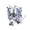


 PDBj
PDBj


 Assembly


 Mass: 407.505 Da / Num. of mol.: 1 / Source method: obtained synthetically / Formula: C24H29N3O3
 Mass: 18.015 Da / Num. of mol.: 3 / Source method: isolated from a natural source / Formula: H2O
 Sample preparation
 Processing