 Movie
Movie Controller


+ Open data

- Basic information
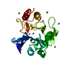
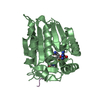
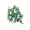
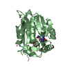
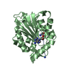
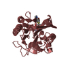
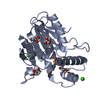
| Entry | Database: PDB / ID: 6pvb | |||||||||
|---|---|---|---|---|---|---|---|---|---|---|
| Title | The structure of NTMT1 in complex with compound 6 | |||||||||
 Components Components |
| |||||||||
 Keywords Keywords | transferase/transferase inhibitor / methyltransferase / enzyme / inhibitor complex / TRANSFERASE / transferase-transferase inhibitor complex | |||||||||
| Function / homology |  Function and homology information Function and homology informationN-terminal peptidyl-glycine methylation / N-terminal peptidyl-serine dimethylation / N-terminal peptidyl-serine trimethylation / N-terminal peptidyl-proline dimethylation / protein N-terminal methyltransferase / N-terminal protein N-methyltransferase activity / protein methyltransferase activity / spindle organization / histone methyltransferase activity / chromosome segregation ...N-terminal peptidyl-glycine methylation / N-terminal peptidyl-serine dimethylation / N-terminal peptidyl-serine trimethylation / N-terminal peptidyl-proline dimethylation / protein N-terminal methyltransferase / N-terminal protein N-methyltransferase activity / protein methyltransferase activity / spindle organization / histone methyltransferase activity / chromosome segregation / nucleoplasm / nucleus / cytosol / cytoplasm Similarity search - Function | |||||||||
| Biological species |  Homo sapiens (human) Homo sapiens (human)synthetic construct (others) | |||||||||
| Method |  X-RAY DIFFRACTION / SYNCHROTRON / MOLECULAR REPLACEMENT / Resolution: 1.5 Å X-RAY DIFFRACTION / SYNCHROTRON / MOLECULAR REPLACEMENT / Resolution: 1.5 Å | |||||||||
 Authors Authors | Noinaj, N. / Chen, D. / Huang, R. | |||||||||
| Funding support |  United States, 2items United States, 2items
| |||||||||
 Citation Citation | Journal: J.Med.Chem. / Year: 2020 Title: Probing the Plasticity in the Active Site of Protein N-terminal Methyltransferase 1 Using Bisubstrate Analogues. Authors: Chen, D. / Dong, C. / Dong, G. / Srinivasan, K. / Min, J. / Noinaj, N. / Huang, R. | |||||||||
| History |
|
- Structure visualization
| Structure viewer | Molecule: MolmilJmol/JSmol |
|---|
- Downloads & links
-Download
| PDBx/mmCIF format | 6pvb.cif.gz | 69.8 KB | Display | PDBx/mmCIF format |
|---|---|---|---|---|
| PDB format | pdb6pvb.ent.gz | 47.1 KB | Display | PDB format |
| PDBx/mmJSON format | 6pvb.json.gz | Tree view | PDBx/mmJSON format | |
| Others |  Other downloads Other downloads |
-Validation report
| Arichive directory | https://data.pdbj.org/pub/pdb/validation_reports/pv/6pvbftp://data.pdbj.org/pub/pdb/validation_reports/pv/6pvb | HTTPS FTP |
|---|
-Related structure data
| Related structure data |  6wj7C 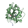 6dtnS S: Starting model for refinement C: citing same article ( |
|---|---|
| Similar structure data |


-Links
 PDBj
PDBj- Assembly
| Deposited unit | 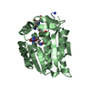
| ||||||||
|---|---|---|---|---|---|---|---|---|---|
| 1 |
| ||||||||
| Unit cell |
|
-Components
| #1: Protein | Mass: 27320.074 Da / Num. of mol.: 1 Source method: isolated from a genetically manipulated source Source: (gene. exp.) Homo sapiens (human) / Gene: NTMT1, C9orf32, METTL11A, NRMT, NRMT1, AD-003 / Production host:  References: UniProt: Q9BV86, protein N-terminal methyltransferase |
|---|---|
| #2: Protein/peptide | Mass: 1044.276 Da / Num. of mol.: 1 / Source method: obtained synthetically / Source: (synth.) synthetic construct (others) |
| #3: Chemical | ChemComp-SAH /   Type: L-peptide linking / Mass: 384.411 Da / Num. of mol.: 1 / Source method: isolated from a natural source / Formula: C14H20N6O5S Type: L-peptide linking / Mass: 384.411 Da / Num. of mol.: 1 / Source method: isolated from a natural source / Formula: C14H20N6O5S |
| #4: Water | ChemComp-HOH /  Mass: 18.015 Da / Num. of mol.: 163 / Source method: isolated from a natural source / Formula: H2O Mass: 18.015 Da / Num. of mol.: 163 / Source method: isolated from a natural source / Formula: H2O |
| Has ligand of interest | Y |
-Experimental details
-Experiment
| Experiment | Method: X-RAY DIFFRACTION / Number of used crystals: 1 |
|---|
- Sample preparation
| Crystal | Density Matthews: 2.31 Å3/Da / Density % sol: 46.7 % |
|---|---|
| Crystal grow | Temperature: 293 K / Method: vapor diffusion, hanging drop / Details: 1.5 M lithium sulfate, 0.1 M sodium HEPES 7.5 |
-Data collection
| Diffraction | Mean temperature: 100 K / Serial crystal experiment: N |
|---|---|
| Diffraction source | Source: SYNCHROTRON / Site: APS / Beamline: 23-ID-D / Wavelength: 1.0332 Å |
| Detector | Type: DECTRIS PILATUS3 6M / Detector: PIXEL / Date: Jun 7, 2018 |
| Radiation | Protocol: SINGLE WAVELENGTH / Monochromatic (M) / Laue (L): M / Scattering type: x-ray |
| Radiation wavelength | Wavelength: 1.0332 Å / Relative weight: 1 |
| Reflection | Resolution: 1.5→30 Å / Num. obs: 41225 / % possible obs: 99.8 % / Redundancy: 5.3 % / Biso Wilson estimate: 28.86 Å2 / CC1/2: 1 / Rsym value: 0.05 / Net I/σ(I): 14.5 |
| Reflection shell | Resolution: 1.5→1.53 Å / Redundancy: 5.3 % / Mean I/σ(I) obs: 0.4 / Num. unique obs: 2962 / CC1/2: 0.17 / Rsym value: 3.3 / % possible all: 98.3 |
- Processing
| Software |
| |||||||||||||||||||||||||||||||||||||||||||||||||||||||||||||||||||||||||||||||||||||||||||||||||||||||||
|---|---|---|---|---|---|---|---|---|---|---|---|---|---|---|---|---|---|---|---|---|---|---|---|---|---|---|---|---|---|---|---|---|---|---|---|---|---|---|---|---|---|---|---|---|---|---|---|---|---|---|---|---|---|---|---|---|---|---|---|---|---|---|---|---|---|---|---|---|---|---|---|---|---|---|---|---|---|---|---|---|---|---|---|---|---|---|---|---|---|---|---|---|---|---|---|---|---|---|---|---|---|---|---|---|---|---|
| Refinement | Method to determine structure: MOLECULAR REPLACEMENT Starting model: 6DTN Resolution: 1.5→29.48 Å / SU ML: 0.25 / Cross valid method: THROUGHOUT / σ(F): 1.33 / Phase error: 29.19 / Stereochemistry target values: ML
| |||||||||||||||||||||||||||||||||||||||||||||||||||||||||||||||||||||||||||||||||||||||||||||||||||||||||
| Solvent computation | Shrinkage radii: 0.9 Å / VDW probe radii: 1.11 Å / Solvent model: FLAT BULK SOLVENT MODEL | |||||||||||||||||||||||||||||||||||||||||||||||||||||||||||||||||||||||||||||||||||||||||||||||||||||||||
| Displacement parameters | Biso max: 94.95 Å2 / Biso mean: 38.2219 Å2 / Biso min: 20.04 Å2 | |||||||||||||||||||||||||||||||||||||||||||||||||||||||||||||||||||||||||||||||||||||||||||||||||||||||||
| Refinement step | Cycle: final / Resolution: 1.5→29.48 Å
| |||||||||||||||||||||||||||||||||||||||||||||||||||||||||||||||||||||||||||||||||||||||||||||||||||||||||
| LS refinement shell | Refine-ID: X-RAY DIFFRACTION / Rfactor Rfree error: 0 / Total num. of bins used: 14
|
