 Movie
Movie Controller


+ Open data

- Basic information
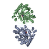
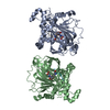
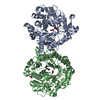
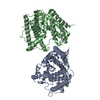
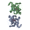
| Entry | Database: PDB / ID: 6prm | ||||||
|---|---|---|---|---|---|---|---|
| Title | Crystal structure of apo PsS1_19B | ||||||
 Components Components | exo-4S-kappa carrageenan S1 sulfatase | ||||||
 Keywords Keywords | HYDROLASE / S1 sulfatase | ||||||
| Function / homology | sulfuric ester hydrolase activity / Sulfatases signature 1. / Sulfatase, conserved site / Sulfatase, N-terminal / Sulfatase / Alkaline-phosphatase-like, core domain superfamily / Sulfatase Function and homology information Function and homology information | ||||||
| Biological species |  Pseudoalteromonas fuliginea (bacteria) Pseudoalteromonas fuliginea (bacteria) | ||||||
| Method |  X-RAY DIFFRACTION / SYNCHROTRON / MOLECULAR REPLACEMENT / molecular replacement / Resolution: 2.5 Å X-RAY DIFFRACTION / SYNCHROTRON / MOLECULAR REPLACEMENT / molecular replacement / Resolution: 2.5 Å | ||||||
 Authors Authors | Hettle, A.G. / Boraston, A.B. | ||||||
| Funding support |  Canada, 1items Canada, 1items
| ||||||
 Citation Citation | Journal: Commun Biol / Year: 2019 Title: Insights into the kappa / iota-carrageenan metabolism pathway of some marinePseudoalteromonasspecies. Authors: Hettle, A.G. / Hobbs, J.K. / Pluvinage, B. / Vickers, C. / Abe, K.T. / Salama-Alber, O. / McGuire, B.E. / Hehemann, J.H. / Hui, J.P.M. / Berrue, F. / Banskota, A. / Zhang, J. / Bottos, E.M. ...Authors: Hettle, A.G. / Hobbs, J.K. / Pluvinage, B. / Vickers, C. / Abe, K.T. / Salama-Alber, O. / McGuire, B.E. / Hehemann, J.H. / Hui, J.P.M. / Berrue, F. / Banskota, A. / Zhang, J. / Bottos, E.M. / Van Hamme, J. / Boraston, A.B. | ||||||
| History |
|
- Structure visualization
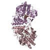
| Structure viewer | Molecule: MolmilJmol/JSmol |
|---|
- Downloads & links
-Download
| PDBx/mmCIF format | 6prm.cif.gz | 352.8 KB | Display | PDBx/mmCIF format |
|---|---|---|---|---|
| PDB format | pdb6prm.ent.gz | 284.2 KB | Display | PDB format |
| PDBx/mmJSON format | 6prm.json.gz | Tree view | PDBx/mmJSON format | |
| Others |  Other downloads Other downloads |
-Validation report
| Arichive directory | https://data.pdbj.org/pub/pdb/validation_reports/pr/6prmftp://data.pdbj.org/pub/pdb/validation_reports/pr/6prm | HTTPS FTP |
|---|
-Related structure data
| Related structure data | 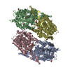 6pnuC 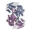 6popC  6psmC 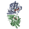 6psoC  6pt4C 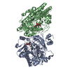 6pt6C  6pt9C  6ptkC  6ptmC 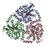 6biaS S: Starting model for refinement C: citing same article ( |
|---|---|
| Similar structure data |


-Links
 PDBj
PDBj
- Assembly
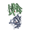
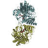
| Deposited unit | 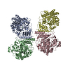
| ||||||||||||||||||||||||||||||||||||||||||||||||||||||||||||||||||||||||||||||||||||||||||||||||||||||||||||||||||||||||||||||||||||||||||||||||||||||||||||||||||||||||||||||||
|---|---|---|---|---|---|---|---|---|---|---|---|---|---|---|---|---|---|---|---|---|---|---|---|---|---|---|---|---|---|---|---|---|---|---|---|---|---|---|---|---|---|---|---|---|---|---|---|---|---|---|---|---|---|---|---|---|---|---|---|---|---|---|---|---|---|---|---|---|---|---|---|---|---|---|---|---|---|---|---|---|---|---|---|---|---|---|---|---|---|---|---|---|---|---|---|---|---|---|---|---|---|---|---|---|---|---|---|---|---|---|---|---|---|---|---|---|---|---|---|---|---|---|---|---|---|---|---|---|---|---|---|---|---|---|---|---|---|---|---|---|---|---|---|---|---|---|---|---|---|---|---|---|---|---|---|---|---|---|---|---|---|---|---|---|---|---|---|---|---|---|---|---|---|---|---|---|---|
| 1 | 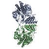
| ||||||||||||||||||||||||||||||||||||||||||||||||||||||||||||||||||||||||||||||||||||||||||||||||||||||||||||||||||||||||||||||||||||||||||||||||||||||||||||||||||||||||||||||||
| 2 | 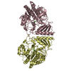
| ||||||||||||||||||||||||||||||||||||||||||||||||||||||||||||||||||||||||||||||||||||||||||||||||||||||||||||||||||||||||||||||||||||||||||||||||||||||||||||||||||||||||||||||||
| Unit cell |
| ||||||||||||||||||||||||||||||||||||||||||||||||||||||||||||||||||||||||||||||||||||||||||||||||||||||||||||||||||||||||||||||||||||||||||||||||||||||||||||||||||||||||||||||||
| Noncrystallographic symmetry (NCS) | NCS domain:
NCS domain segments: Component-ID: _ / Refine code: _
NCS ensembles :
|
-Components
| #1: Protein | Mass: 53067.652 Da / Num. of mol.: 4 Source method: isolated from a genetically manipulated source Source: (gene. exp.) Pseudoalteromonas fuliginea (bacteria) / Strain: PS47 / Gene: EU509_08890 / Production host: #2: Chemical | ChemComp-CA /   Mass: 40.078 Da / Num. of mol.: 4 / Source method: obtained synthetically / Formula: Ca Mass: 40.078 Da / Num. of mol.: 4 / Source method: obtained synthetically / Formula: Ca#3: Water | ChemComp-HOH / |  Mass: 18.015 Da / Num. of mol.: 104 / Source method: isolated from a natural source / Formula: H2O Mass: 18.015 Da / Num. of mol.: 104 / Source method: isolated from a natural source / Formula: H2OHas ligand of interest | N | |
|---|
-Experimental details
-Experiment
| Experiment | Method: X-RAY DIFFRACTION / Number of used crystals: 1 |
|---|
- Sample preparation
| Crystal | Density Matthews: 2.2 Å3/Da / Density % sol: 44.11 % |
|---|---|
| Crystal grow | Temperature: 291 K / Method: vapor diffusion, sitting drop / Details: PEG 3350, sodium acetate trihydrate |
-Data collection
| Diffraction | Mean temperature: 100 K / Serial crystal experiment: N |
|---|---|
| Diffraction source | Source: SYNCHROTRON / Site: CLSI / Beamline: 08ID-1 / Wavelength: 0.97949 Å |
| Detector | Type: RAYONIX MX-300 / Detector: CCD / Date: Dec 5, 2013 |
| Radiation | Protocol: SINGLE WAVELENGTH / Monochromatic (M) / Laue (L): M / Scattering type: x-ray |
| Radiation wavelength | Wavelength: 0.97949 Å / Relative weight: 1 |
| Reflection | Resolution: 2.5→104.2 Å / Num. obs: 58432 / % possible obs: 97.9 % / Redundancy: 3.3 % / Rmerge(I) obs: 0.108 / Net I/σ(I): 8.4 |
| Reflection shell | Resolution: 2.5→2.57 Å / Rmerge(I) obs: 0.334 / Num. unique obs: 4508 |
-Phasing
| Phasing | Method: molecular replacement |
|---|
- Processing
| Software |
| |||||||||||||||||||||||||||||||||||||||||||||||||||||||||||||||||
|---|---|---|---|---|---|---|---|---|---|---|---|---|---|---|---|---|---|---|---|---|---|---|---|---|---|---|---|---|---|---|---|---|---|---|---|---|---|---|---|---|---|---|---|---|---|---|---|---|---|---|---|---|---|---|---|---|---|---|---|---|---|---|---|---|---|---|
| Refinement | Method to determine structure: MOLECULAR REPLACEMENT Starting model: 6BIA Resolution: 2.5→47.6 Å / Cor.coef. Fo:Fc: 0.895 / Cor.coef. Fo:Fc free: 0.866 / SU B: 13.753 / SU ML: 0.303 / Cross valid method: THROUGHOUT / σ(F): 0 / ESU R Free: 0.353 Details: HYDROGENS HAVE BEEN ADDED IN THE RIDING POSITIONS U VALUES : REFINED INDIVIDUALLY
| |||||||||||||||||||||||||||||||||||||||||||||||||||||||||||||||||
| Solvent computation | Ion probe radii: 0.8 Å / Shrinkage radii: 0.8 Å / VDW probe radii: 1.2 Å | |||||||||||||||||||||||||||||||||||||||||||||||||||||||||||||||||
| Displacement parameters | Biso max: 92.88 Å2 / Biso mean: 30.899 Å2 / Biso min: 4.02 Å2
| |||||||||||||||||||||||||||||||||||||||||||||||||||||||||||||||||
| Refinement step | Cycle: final / Resolution: 2.5→47.6 Å
| |||||||||||||||||||||||||||||||||||||||||||||||||||||||||||||||||
| Refine LS restraints |
| |||||||||||||||||||||||||||||||||||||||||||||||||||||||||||||||||
| Refine LS restraints NCS | Refine-ID: X-RAY DIFFRACTION / Type: interatomic distance / Weight position: 0.05
| |||||||||||||||||||||||||||||||||||||||||||||||||||||||||||||||||
| LS refinement shell | Resolution: 2.5→2.565 Å / Rfactor Rfree error: 0 / Total num. of bins used: 20
|
