 Movie
Movie Controller


+ Open data

- Basic information
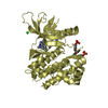
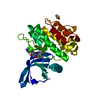
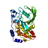
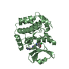
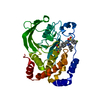
| Entry | Database: PDB / ID: 6pq9 | ||||||
|---|---|---|---|---|---|---|---|
| Title | Crystal Structure of TLA-1 S70G extended spectrum Beta-lactamase | ||||||
 Components Components | Beta-lactamase | ||||||
 Keywords Keywords | HYDROLASE / Lactamase / Antibiotic / Resistance / HIDROLASE | ||||||
| Function / homology |  Function and homology information Function and homology informationbeta-lactam antibiotic catabolic process / beta-lactamase activity / beta-lactamase / response to antibiotic Similarity search - Function | ||||||
| Biological species |  | ||||||
| Method |  X-RAY DIFFRACTION / SYNCHROTRON / MOLECULAR REPLACEMENT / molecular replacement / Resolution: 2.191 Å X-RAY DIFFRACTION / SYNCHROTRON / MOLECULAR REPLACEMENT / molecular replacement / Resolution: 2.191 Å | ||||||
 Authors Authors | Rudino-Pinera, E. / Cifuentes-Castro, V.H. / Rodriguez-Alamazan, C. | ||||||
 Citation Citation | Journal: Biochem.Biophys.Res.Commun. / Year: 2020 Title: The crystal structure of ESBL TLA-1 in complex with clavulanic acid reveals a second acylation site. Authors: Cifuentes-Castro, V. / Rodriguez-Almazan, C. / Silva-Sanchez, J. / Rudino-Pinera, E. | ||||||
| History |
|
- Structure visualization
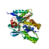
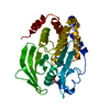
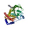
| Structure viewer | Molecule: MolmilJmol/JSmol |
|---|
- Downloads & links
-Download
| PDBx/mmCIF format | 6pq9.cif.gz | 76.8 KB | Display | PDBx/mmCIF format |
|---|---|---|---|---|
| PDB format | pdb6pq9.ent.gz | 54.5 KB | Display | PDB format |
| PDBx/mmJSON format | 6pq9.json.gz | Tree view | PDBx/mmJSON format | |
| Others |  Other downloads Other downloads |
-Validation report
| Arichive directory | https://data.pdbj.org/pub/pdb/validation_reports/pq/6pq9ftp://data.pdbj.org/pub/pdb/validation_reports/pq/6pq9 | HTTPS FTP |
|---|
-Related structure data
| Related structure data |  6nvtC  6nvuSC 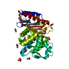 6pq8C S: Starting model for refinement C: citing same article ( |
|---|---|
| Similar structure data |


-Links
 PDBj
PDBj


- Assembly
| Deposited unit | 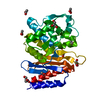
| ||||||||
|---|---|---|---|---|---|---|---|---|---|
| 1 |
| ||||||||
| Unit cell |
|
-Components
-Protein , 1 types, 1 molecules A
| #1: Protein | Mass: 33342.285 Da / Num. of mol.: 1 / Mutation: S70G Source method: isolated from a genetically manipulated source Source: (gene. exp.) |
|---|
-Non-polymers , 5 types, 196 molecules 


| #2: Chemical | ChemComp-ASP /  Type: L-peptide linking / Mass: 133.103 Da / Num. of mol.: 1 / Source method: obtained synthetically / Formula: C4H7NO4 Type: L-peptide linking / Mass: 133.103 Da / Num. of mol.: 1 / Source method: obtained synthetically / Formula: C4H7NO4 | ||||||
|---|---|---|---|---|---|---|---|
| #3: Chemical | ChemComp-ACT /  Mass: 59.044 Da / Num. of mol.: 7 / Source method: obtained synthetically / Formula: C2H3O2 Mass: 59.044 Da / Num. of mol.: 7 / Source method: obtained synthetically / Formula: C2H3O2#4: Chemical |  Mass: 92.094 Da / Num. of mol.: 2 / Source method: obtained synthetically / Formula: C3H8O3 Mass: 92.094 Da / Num. of mol.: 2 / Source method: obtained synthetically / Formula: C3H8O3#5: Chemical | ChemComp-SO4 / |  Mass: 96.063 Da / Num. of mol.: 1 / Source method: obtained synthetically / Formula: SO4 Mass: 96.063 Da / Num. of mol.: 1 / Source method: obtained synthetically / Formula: SO4#6: Water | ChemComp-HOH / | Mass: 18.015 Da / Num. of mol.: 185 / Source method: isolated from a natural source / Formula: H2O |
-Details
| Has ligand of interest | N |
|---|
-Experimental details
-Experiment
| Experiment | Method: X-RAY DIFFRACTION / Number of used crystals: 1 |
|---|
- Sample preparation
| Crystal | Density Matthews: 3.97 Å3/Da / Density % sol: 69.05 % |
|---|---|
| Crystal grow | Temperature: 291.15 K / Method: vapor diffusion, sitting drop / pH: 4.5 Details: 2.4 M Ammonium Sulfate, 100 mM Sodium Phosphate dibasic/ Citric acid pH 4.5 |
-Data collection
| Diffraction | Mean temperature: 100 K / Serial crystal experiment: N |
|---|---|
| Diffraction source | Source: SYNCHROTRON / Site: APS  / Beamline: 19-BM / Wavelength: 0.9793326 Å / Beamline: 19-BM / Wavelength: 0.9793326 Å |
| Detector | Type: ADSC QUANTUM 210r / Detector: CCD / Date: Oct 17, 2018 / Details: Sagitally focusing 2nd crystal |
| Radiation | Monochromator: Si (111) Rosenbaum-Rock double-crystal monochromator Protocol: SINGLE WAVELENGTH / Monochromatic (M) / Laue (L): M / Scattering type: x-ray |
| Radiation wavelength | Wavelength: 0.9793326 Å / Relative weight: 1 |
| Reflection | Resolution: 2.191→19.89 Å / Num. obs: 26170 / % possible obs: 99.9 % / Redundancy: 10.74 % / Biso Wilson estimate: 41.54 Å2 / CC1/2: 1 / Rmerge(I) obs: 0.082 / Rrim(I) all: 0.084 / Rsym value: 0.084 / Net I/σ(I): 30.14 |
| Reflection shell | Resolution: 2.2→2.3 Å / Rmerge(I) obs: 1.12 / Num. unique obs: 3202 / CC1/2: 0.898 / Rrim(I) all: 1.15 / Rsym value: 1.16 / % possible all: 100 |
-Phasing
| Phasing | Method: molecular replacement |
|---|
- Processing
| Software |
| ||||||||||||||||||||||||||||||||||||||||||||||||||||||||||||
|---|---|---|---|---|---|---|---|---|---|---|---|---|---|---|---|---|---|---|---|---|---|---|---|---|---|---|---|---|---|---|---|---|---|---|---|---|---|---|---|---|---|---|---|---|---|---|---|---|---|---|---|---|---|---|---|---|---|---|---|---|---|
| Refinement | Method to determine structure: MOLECULAR REPLACEMENT Starting model: 6NVU Resolution: 2.191→19.89 Å / SU ML: 0.32 / Cross valid method: THROUGHOUT / σ(F): 1.36 / Phase error: 25.42
| ||||||||||||||||||||||||||||||||||||||||||||||||||||||||||||
| Solvent computation | Shrinkage radii: 0.9 Å / VDW probe radii: 1.11 Å | ||||||||||||||||||||||||||||||||||||||||||||||||||||||||||||
| Displacement parameters | Biso max: 104.89 Å2 / Biso mean: 44.1204 Å2 / Biso min: 25.64 Å2 | ||||||||||||||||||||||||||||||||||||||||||||||||||||||||||||
| Refinement step | Cycle: final / Resolution: 2.191→19.89 Å
| ||||||||||||||||||||||||||||||||||||||||||||||||||||||||||||
| Refine LS restraints |
| ||||||||||||||||||||||||||||||||||||||||||||||||||||||||||||
| LS refinement shell | Refine-ID: X-RAY DIFFRACTION / Rfactor Rfree error: 0
|
