| Entry | Database: PDB / ID: 5ajq
|
|---|
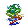
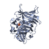
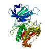
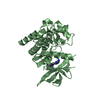
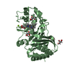
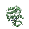
| Title | Human LOK (STK10) in complex with Bosutinib |
|---|
 Components Components | SERINE/THREONINE-PROTEIN KINASE 10 |
|---|
 Keywords Keywords | TRANSFERASE |
|---|
| Function / homology |  Function and homology information Function and homology information
lymphocyte aggregation / regulation of lymphocyte migration / RHOB GTPase cycle / RHOC GTPase cycle / RHOA GTPase cycle / specific granule membrane / protein autophosphorylation / protein phosphorylation / non-specific serine/threonine protein kinase / intracellular signal transduction ...lymphocyte aggregation / regulation of lymphocyte migration / RHOB GTPase cycle / RHOC GTPase cycle / RHOA GTPase cycle / specific granule membrane / protein autophosphorylation / protein phosphorylation / non-specific serine/threonine protein kinase / intracellular signal transduction / nuclear body / protein serine kinase activity / protein serine/threonine kinase activity / Neutrophil degranulation / protein homodimerization activity / extracellular exosome / nucleoplasm / ATP binding / identical protein binding / plasma membrane / cytoplasm / cytosolSimilarity search - Function Serine/threonine-protein kinase 10, catalytic domain / Polo kinase kinase / : / Polo kinase kinase / Phosphorylase Kinase; domain 1 / Phosphorylase Kinase; domain 1 / Transferase(Phosphotransferase) domain 1 / Transferase(Phosphotransferase); domain 1 / Serine/threonine-protein kinase, active site / Serine/Threonine protein kinases active-site signature. ...Serine/threonine-protein kinase 10, catalytic domain / Polo kinase kinase / : / Polo kinase kinase / Phosphorylase Kinase; domain 1 / Phosphorylase Kinase; domain 1 / Transferase(Phosphotransferase) domain 1 / Transferase(Phosphotransferase); domain 1 / Serine/threonine-protein kinase, active site / Serine/Threonine protein kinases active-site signature. / Protein kinase domain / Serine/Threonine protein kinases, catalytic domain / Protein kinase, ATP binding site / Protein kinases ATP-binding region signature. / Protein kinase domain profile. / Protein kinase domain / Protein kinase-like domain superfamily / 2-Layer Sandwich / Orthogonal Bundle / Mainly Alpha / Alpha BetaSimilarity search - Domain/homology |
|---|
| Biological species |  HOMO SAPIENS (human) HOMO SAPIENS (human) |
|---|
| Method |  X-RAY DIFFRACTION / SYNCHROTRON / MOLECULAR REPLACEMENT / Resolution: 2.2 Å X-RAY DIFFRACTION / SYNCHROTRON / MOLECULAR REPLACEMENT / Resolution: 2.2 Å |
|---|
 Authors Authors | Elkins, J.M. / Salah, E. / Pinkas, D.M. / Krojer, T. / Kopec, J. / Bountra, C. / Edwards, A.M. / Knapp, S. |
|---|
 Citation Citation | Journal: To be Published
Title: Stk10 with Bosutinib
Authors: Elkins, J.M. / Salah, E. / Knapp, S. |
|---|
| History | | Deposition | Feb 26, 2015 | Deposition site: PDBE / Processing site: PDBE |
|---|
| Revision 1.0 | Mar 11, 2015 | Provider: repository / Type: Initial release |
|---|
| Revision 1.1 | Apr 8, 2015 | Group: Structure summary |
|---|
| Revision 1.2 | Oct 23, 2024 | Group: Data collection / Database references ...Data collection / Database references / Derived calculations / Other / Refinement description / Structure summary
Category: chem_comp_atom / chem_comp_bond ...chem_comp_atom / chem_comp_bond / database_2 / pdbx_database_status / pdbx_entry_details / pdbx_modification_feature / struct_ncs_dom_lim / struct_site
Item: _database_2.pdbx_DOI / _database_2.pdbx_database_accession ..._database_2.pdbx_DOI / _database_2.pdbx_database_accession / _pdbx_database_status.status_code_sf / _struct_ncs_dom_lim.beg_auth_comp_id / _struct_ncs_dom_lim.beg_label_asym_id / _struct_ncs_dom_lim.beg_label_comp_id / _struct_ncs_dom_lim.beg_label_seq_id / _struct_ncs_dom_lim.end_auth_comp_id / _struct_ncs_dom_lim.end_label_asym_id / _struct_ncs_dom_lim.end_label_comp_id / _struct_ncs_dom_lim.end_label_seq_id / _struct_site.pdbx_auth_asym_id / _struct_site.pdbx_auth_comp_id / _struct_site.pdbx_auth_seq_id |
|---|
|
|---|
 Movie
Movie Controller


 Open data
 Basic information
 Structure visualization
 Downloads & links
 Other downloads
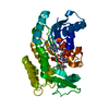


 PDBj
PDBj
 Assembly


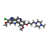
 Mass: 530.446 Da / Num. of mol.: 2 / Source method: obtained synthetically / Formula: C26H29Cl2N5O3 / Comment: inhibitor, medication*YM
 Mass: 35.453 Da / Num. of mol.: 1 / Source method: obtained synthetically / Formula: Cl
 Mass: 62.068 Da / Num. of mol.: 1 / Source method: obtained synthetically / Formula: C2H6O2
 Mass: 18.015 Da / Num. of mol.: 86 / Source method: isolated from a natural source / Formula: H2O
 Sample preparation
 / Beamline: I02 / Wavelength: 0.97949
  Processing