National Institutes of Health/National Institute on Drug Abuse (NIH/NIDA)
R21DA042298
United States
National Institutes of Health/National Institute of General Medical Sciences (NIH/NIGMS)
R01GM124152
United States
Citation
Journal: Structure / Year: 2020 Title: Structure Determination from Lipidic Cubic Phase Embedded Microcrystals by MicroED. Authors: Lan Zhu / Guanhong Bu / Liang Jing / Dan Shi / Ming-Yue Lee / Tamir Gonen / Wei Liu / Brent L Nannenga / Abstract: The lipidic cubic phase (LCP) technique has proved to facilitate the growth of high-quality crystals that are otherwise difficult to grow by other methods. However, the crystal size optimization ...The lipidic cubic phase (LCP) technique has proved to facilitate the growth of high-quality crystals that are otherwise difficult to grow by other methods. However, the crystal size optimization process could be time and resource consuming, if it ever happens. Therefore, improved techniques for structure determination using these small crystals is an important strategy in diffraction technology development. Microcrystal electron diffraction (MicroED) is a technique that uses a cryo-transmission electron microscopy to collect electron diffraction data and determine high-resolution structures from very thin micro- and nanocrystals. In this work, we have used modified LCP and MicroED protocols to analyze crystals embedded in LCP converted by 2-methyl-2,4-pentanediol or lipase, including Proteinase K crystals grown in solution, cholesterol crystals, and human adenosine A receptor crystals grown in LCP. These results set the stage for the use of MicroED to analyze microcrystalline samples grown in LCP, especially for those highly challenging membrane protein targets.
In the structure databanks used in Yorodumi, some data are registered as the other names, "COVID-19 virus" and "2019-nCoV". Here are the details of the virus and the list of structure data.
Jan 31, 2019. EMDB accession codes are about to change! (news from PDBe EMDB page)
EMDB accession codes are about to change! (news from PDBe EMDB page)
The allocation of 4 digits for EMDB accession codes will soon come to an end. Whilst these codes will remain in use, new EMDB accession codes will include an additional digit and will expand incrementally as the available range of codes is exhausted. The current 4-digit format prefixed with “EMD-” (i.e. EMD-XXXX) will advance to a 5-digit format (i.e. EMD-XXXXX), and so on. It is currently estimated that the 4-digit codes will be depleted around Spring 2019, at which point the 5-digit format will come into force.
The EM Navigator/Yorodumi systems omit the EMD- prefix.
Related info.:Q: What is EMD? / ID/Accession-code notation in Yorodumi/EM Navigator
Yorodumi is a browser for structure data from EMDB, PDB, SASBDB, etc.
This page is also the successor to EM Navigator detail page, and also detail information page/front-end page for Omokage search.
The word "yorodu" (or yorozu) is an old Japanese word meaning "ten thousand". "mi" (miru) is to see.
Related info.:EMDB / PDB / SASBDB / Comparison of 3 databanks / Yorodumi Search / Aug 31, 2016. New EM Navigator & Yorodumi / Yorodumi Papers / Jmol/JSmol / Function and homology information / Changes in new EM Navigator and Yorodumi
 Movie
Movie Controller


 Open data
 Basic information
 Components
 Keywords
 Function and homology information
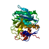
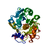
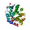
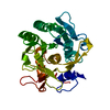
 Parengyodontium album (fungus)
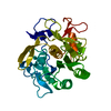
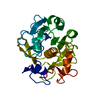
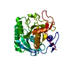
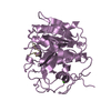
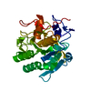
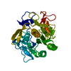
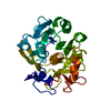
 MOLECULAR REPLACEMENT / cryo EM / Resolution: 2 Å
  Authors
 United States, 4items
  Citation
 Structure visualization
 Downloads & links
 Other downloads


 PDBj
PDBj

 Assembly
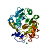

 Mass: 40.078 Da / Num. of mol.: 2 / Source method: obtained synthetically / Formula: Ca / Feature type: SUBJECT OF INVESTIGATION
 Mass: 62.005 Da / Num. of mol.: 1 / Source method: obtained synthetically / Formula: NO3
 Mass: 18.015 Da / Num. of mol.: 95 / Source method: isolated from a natural source / Formula: H2O
 Sample preparation
 Processing