 Movie
Movie Controller

[English] 日本語
 Yorodumi
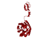
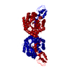
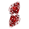
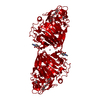
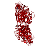
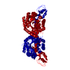
- PDB-6ppx: Crystal structure of metal-free NeuB, an N-acetylneuraminate synt... -
+ Open data

- Basic information
| Entry | Database: PDB / ID: 6ppx | |||||||||
|---|---|---|---|---|---|---|---|---|---|---|
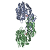
| Title | Crystal structure of metal-free NeuB, an N-acetylneuraminate synthase from Neisseria meningitidis in complex with malate | |||||||||
 Components Components | N-acetylneuraminate synthase | |||||||||
 Keywords Keywords | BIOSYNTHETIC PROTEIN / TRANSFERASE / NeuB / sialic acid synthase / N-acetylneuraminate synthase | |||||||||
| Function / homology |  Function and homology information Function and homology informationN-acetylneuraminate synthase / N-acetylneuraminate synthase activity / N-acylneuraminate-9-phosphate synthase activity / carbohydrate biosynthetic process / metal ion binding Similarity search - Function | |||||||||
| Biological species |  Neisseria meningitidis serogroup B (bacteria) Neisseria meningitidis serogroup B (bacteria) | |||||||||
| Method |  X-RAY DIFFRACTION / MOLECULAR REPLACEMENT / Resolution: 1.85 Å X-RAY DIFFRACTION / MOLECULAR REPLACEMENT / Resolution: 1.85 Å | |||||||||
 Authors Authors | Rosanally, A.Z. / Junop, M.S. / Berti, P.J. | |||||||||
| Funding support |  Canada, 2items Canada, 2items
| |||||||||
 Citation Citation | Journal: Biochemistry / Year: 2019 Title: NeuNAc Oxime: A Slow-Binding and Effectively Irreversible Inhibitor of the Sialic Acid Synthase NeuB. Authors: Popovic, V. / Morrison, E. / Rosanally, A.Z. / Balachandran, N. / Senson, A.W. / Szabla, R. / Junop, M.S. / Berti, P.J. | |||||||||
| History |
|
- Structure visualization
| Structure viewer | Molecule: MolmilJmol/JSmol |
|---|
- Downloads & links
-Download
| PDBx/mmCIF format | 6ppx.cif.gz | 96.3 KB | Display | PDBx/mmCIF format |
|---|---|---|---|---|
| PDB format | pdb6ppx.ent.gz | 64.5 KB | Display | PDB format |
| PDBx/mmJSON format | 6ppx.json.gz | Tree view | PDBx/mmJSON format | |
| Others |  Other downloads Other downloads |
-Validation report
| Arichive directory | https://data.pdbj.org/pub/pdb/validation_reports/pp/6ppxftp://data.pdbj.org/pub/pdb/validation_reports/pp/6ppx | HTTPS FTP |
|---|
-Related structure data
| Related structure data | 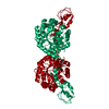 6ppwC  6ppyC  6ppzC 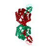 1xuzS S: Starting model for refinement C: citing same article ( |
|---|---|
| Similar structure data |


-Links
 PDBj
PDBj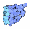


- Assembly
| Deposited unit | 
| ||||||||||||
|---|---|---|---|---|---|---|---|---|---|---|---|---|---|
| 1 | 
| ||||||||||||
| Unit cell |
|
-Components
| #1: Protein | Mass: 38395.730 Da / Num. of mol.: 1 Source method: isolated from a genetically manipulated source Source: (gene. exp.) Neisseria meningitidis serogroup B (bacteria)Gene: synC / Production host: |
|---|---|
| #2: Chemical | ChemComp-MLT /   Mass: 134.087 Da / Num. of mol.: 1 / Source method: obtained synthetically / Formula: C4H6O5 Mass: 134.087 Da / Num. of mol.: 1 / Source method: obtained synthetically / Formula: C4H6O5 |
| #3: Water | ChemComp-HOH /  Mass: 18.015 Da / Num. of mol.: 339 / Source method: isolated from a natural source / Formula: H2O Mass: 18.015 Da / Num. of mol.: 339 / Source method: isolated from a natural source / Formula: H2O |
| Has ligand of interest | N |
-Experimental details
-Experiment
| Experiment | Method: X-RAY DIFFRACTION / Number of used crystals: 1 |
|---|
- Sample preparation
| Crystal | Density Matthews: 2.23 Å3/Da / Density % sol: 44.95 % |
|---|---|
| Crystal grow | Temperature: 291 K / Method: vapor diffusion, hanging drop / pH: 6.2 Details: 3% (v/v) MPD 1.75M Malic acid 10mM magnesium chloride 10mM N-acetylneuraminic acid |
-Data collection
| Diffraction | Mean temperature: 100 K / Serial crystal experiment: N |
|---|---|
| Diffraction source | Source: ROTATING ANODE / Type: RIGAKU RU300 / Wavelength: 1.5418 Å |
| Detector | Type: RIGAKU RAXIS IV++ / Detector: IMAGE PLATE / Date: Jan 29, 2008 |
| Radiation | Monochromator: Yale Mirrors / Protocol: SINGLE WAVELENGTH / Monochromatic (M) / Laue (L): M / Scattering type: x-ray |
| Radiation wavelength | Wavelength: 1.5418 Å / Relative weight: 1 |
| Reflection | Resolution: 1.85→77.62 Å / Num. obs: 29645 / % possible obs: 98.5 % / Redundancy: 3.9 % / Biso Wilson estimate: 23.03 Å2 / CC1/2: 0.998 / Rmerge(I) obs: 0.098 / Rpim(I) all: 0.035 / Rrim(I) all: 0.104 / Net I/σ(I): 2.53 |
| Reflection shell | Resolution: 1.85→1.92 Å / Redundancy: 3.8 % / Rmerge(I) obs: 0.673 / Mean I/σ(I) obs: 3.1 / Num. unique obs: 2676 / CC1/2: 0.915 / Rpim(I) all: 0.248 |
- Processing
| Software |
| ||||||||||||||||||||||||||||||||||||||||||||||||||||||||||||||||||||||||||||||||||||
|---|---|---|---|---|---|---|---|---|---|---|---|---|---|---|---|---|---|---|---|---|---|---|---|---|---|---|---|---|---|---|---|---|---|---|---|---|---|---|---|---|---|---|---|---|---|---|---|---|---|---|---|---|---|---|---|---|---|---|---|---|---|---|---|---|---|---|---|---|---|---|---|---|---|---|---|---|---|---|---|---|---|---|---|---|---|
| Refinement | Method to determine structure: MOLECULAR REPLACEMENT Starting model: 1XUZ Resolution: 1.85→46.48 Å / SU ML: 0.2857 / Cross valid method: FREE R-VALUE / σ(F): 1.35 / Phase error: 25.1387
| ||||||||||||||||||||||||||||||||||||||||||||||||||||||||||||||||||||||||||||||||||||
| Solvent computation | Shrinkage radii: 0.9 Å / VDW probe radii: 1.11 Å | ||||||||||||||||||||||||||||||||||||||||||||||||||||||||||||||||||||||||||||||||||||
| Displacement parameters | Biso mean: 24.7 Å2 | ||||||||||||||||||||||||||||||||||||||||||||||||||||||||||||||||||||||||||||||||||||
| Refinement step | Cycle: LAST / Resolution: 1.85→46.48 Å
| ||||||||||||||||||||||||||||||||||||||||||||||||||||||||||||||||||||||||||||||||||||
| Refine LS restraints |
| ||||||||||||||||||||||||||||||||||||||||||||||||||||||||||||||||||||||||||||||||||||
| LS refinement shell |
|
