 Movie
Movie Controller


+ Open data

- Basic information
| Entry | Database: PDB / ID: 4d5q | ||||||||||||
|---|---|---|---|---|---|---|---|---|---|---|---|---|---|
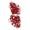
| Title | Hypocrea jecorina Cel7A (wild type) soaked with xylopentaose. | ||||||||||||
 Components Components | CELLULOSE 1,4-BETA-CELLOBIOSIDASE | ||||||||||||
 Keywords Keywords | HYDROLASE / GLYCOSIDE HYDROLASE / CELLOBIOHYDROLASE / CELLULASE. INHIBITION / XYLOOLIGOSACCHARIDES | ||||||||||||
| Function / homology |  Function and homology information Function and homology informationcellulose 1,4-beta-cellobiosidase (non-reducing end) / cellulose 1,4-beta-cellobiosidase activity / cellulose binding / cellulose catabolic process / extracellular region Similarity search - Function | ||||||||||||
| Biological species |  TRICHODERMA REESEI QM9414 (fungus) TRICHODERMA REESEI QM9414 (fungus) | ||||||||||||
| Method |  X-RAY DIFFRACTION / SYNCHROTRON / MOLECULAR REPLACEMENT / Resolution: 1.68 Å X-RAY DIFFRACTION / SYNCHROTRON / MOLECULAR REPLACEMENT / Resolution: 1.68 Å | ||||||||||||
 Authors Authors | Momeni, M.H. / Stahlberg, J. / Hansson, H. | ||||||||||||
 Citation Citation | Journal: FEBS J. / Year: 2015 Title: Structural Insights Into the Inhibition of Cellobiohydrolase Cel7A by Xylooligosaccharides. Authors: Haddad Momeni, M. / Ubhayasekera, W. / Sandgren, M. / Stahlberg, J. / Hansson, H. | ||||||||||||
| History |
|
- Structure visualization
| Structure viewer | Molecule: MolmilJmol/JSmol |
|---|
- Downloads & links
-Download
| PDBx/mmCIF format | 4d5q.cif.gz | 112.7 KB | Display | PDBx/mmCIF format |
|---|---|---|---|---|
| PDB format | pdb4d5q.ent.gz | 85.1 KB | Display | PDB format |
| PDBx/mmJSON format | 4d5q.json.gz | Tree view | PDBx/mmJSON format | |
| Others |  Other downloads Other downloads |
-Validation report
| Arichive directory | https://data.pdbj.org/pub/pdb/validation_reports/d5/4d5qftp://data.pdbj.org/pub/pdb/validation_reports/d5/4d5q | HTTPS FTP |
|---|
-Related structure data
| Related structure data |  4d5iC 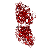 4d5jC  4d5oC 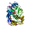 4d5pC 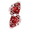 4d5vC  7celS C: citing same article ( S: Starting model for refinement |
|---|---|
| Similar structure data |


-Links
 PDBj
PDBj
- Assembly
| Deposited unit | 
| |||||||||||||||||||||
|---|---|---|---|---|---|---|---|---|---|---|---|---|---|---|---|---|---|---|---|---|---|---|
| 1 | 
| |||||||||||||||||||||
| Unit cell |
| |||||||||||||||||||||
| Components on special symmetry positions |
|
-Components
-Protein , 1 types, 1 molecules A
| #1: Protein | Mass: 46009.719 Da / Num. of mol.: 1 / Fragment: CATALYTIC MODULE, RESIDUES 18-451 Source method: isolated from a genetically manipulated source Details: THE STRUCTURE CONTAINS ONE XYLOTETRAOSE MOLECULE BOUND AT SUBSITES -7 TO -3, A XYLOBIOSE AT -2 TO - 1 AND A XYLOSE MOLECULE AT 1 VIA PARTIAL OCCUPANCY. Source: (gene. exp.) TRICHODERMA REESEI QM9414 (fungus) / Variant: VTT-D-93201 / Plasmid: PEM-F5 / Production host: TRICHODERMA REESEI QM9414 (fungus) / Variant (production host): VTT-D-93201References: UniProt: P62694, cellulose 1,4-beta-cellobiosidase (reducing end) |
|---|
-Sugars , 4 types, 4 molecules 
| #2: Polysaccharide | beta-D-xylopyranose-(1-4)-beta-D-xylopyranose-(1-4)-beta-D-xylopyranose-(1-2)-beta-D-xylopyranose Source method: isolated from a genetically manipulated source |
|---|---|
| #3: Polysaccharide | beta-D-xylopyranose-(1-2)-beta-D-xylopyranose Source method: isolated from a genetically manipulated source |
| #5: Sugar | ChemComp-NAG /  Type: D-saccharide, beta linking / Mass: 221.208 Da / Num. of mol.: 1 Type: D-saccharide, beta linking / Mass: 221.208 Da / Num. of mol.: 1Source method: isolated from a genetically manipulated source Formula: C8H15NO6 |
| #6: Sugar | ChemComp-XYP /  Type: D-saccharide, beta linking / Mass: 150.130 Da / Num. of mol.: 1 Type: D-saccharide, beta linking / Mass: 150.130 Da / Num. of mol.: 1Source method: isolated from a genetically manipulated source Formula: C5H10O5 |
-Non-polymers , 2 types, 561 molecules 

| #4: Chemical |  Mass: 58.933 Da / Num. of mol.: 2 / Source method: obtained synthetically / Formula: Co Mass: 58.933 Da / Num. of mol.: 2 / Source method: obtained synthetically / Formula: Co#7: Water | ChemComp-HOH / | Mass: 18.015 Da / Num. of mol.: 559 / Source method: isolated from a natural source / Formula: H2O |
|---|
-Details
| Has protein modification | Y |
|---|---|
| Nonpolymer details | COBALT (II) ION (CO): 10 MM COBALT CHLORIDE WAS PRESENT IN CRYSTALLISATION SOLUTION PYROGLUTAMIC ...COBALT (II) ION (CO): 10 MM COBALT CHLORIDE WAS PRESENT IN CRYSTALLIS |
-Experimental details
-Experiment
| Experiment | Method: X-RAY DIFFRACTION / Number of used crystals: 3 |
|---|
- Sample preparation
| Crystal | Density Matthews: 2.1 Å3/Da / Density % sol: 41.34 % / Description: NONE |
|---|---|
| Crystal grow | Temperature: 298 K / Method: vapor diffusion, hanging drop / pH: 6 Details: 0.1 M NA-MES (PH 6.0), 20% MONOMETHYL ETHER PEG 5000, 0.01 M COCL2, 12.5% GLYCEROL, VAPOR DIFFUSION, HANGING DROP, TEMPERATURE 298K |
-Data collection
| Diffraction | Mean temperature: 100 K |
|---|---|
| Diffraction source | Source: SYNCHROTRON / Site: MAX II  / Beamline: I911-3 / Wavelength: 1.041 / Beamline: I911-3 / Wavelength: 1.041 |
| Detector | Type: MARRESEARCH MAR165 / Detector: CCD / Date: Jul 17, 2014 / Details: MIRRORS |
| Radiation | Protocol: SINGLE WAVELENGTH / Monochromatic (M) / Laue (L): M / Scattering type: x-ray |
| Radiation wavelength | Wavelength: 1.041 Å / Relative weight: 1 |
| Reflection | Resolution: 1.68→41.64 Å / Num. obs: 42740 / % possible obs: 97.1 % / Observed criterion σ(I): 2 / Redundancy: 4.1 % / Rmerge(I) obs: 0.12 / Net I/σ(I): 8.7 |
| Reflection shell | Resolution: 1.68→1.77 Å / Redundancy: 4 % / Rmerge(I) obs: 0.4 / Mean I/σ(I) obs: 2.8 / % possible all: 99.3 |
- Processing
| Software |
| ||||||||||||||||||||||||||||||||||||||||||||||||||||||||||||||||||||||||||||||||||||||||||||||||||||||||||||||||||||||||||||||||||||||||||||||||||||||||||||||||||||||||||||||||||||||
|---|---|---|---|---|---|---|---|---|---|---|---|---|---|---|---|---|---|---|---|---|---|---|---|---|---|---|---|---|---|---|---|---|---|---|---|---|---|---|---|---|---|---|---|---|---|---|---|---|---|---|---|---|---|---|---|---|---|---|---|---|---|---|---|---|---|---|---|---|---|---|---|---|---|---|---|---|---|---|---|---|---|---|---|---|---|---|---|---|---|---|---|---|---|---|---|---|---|---|---|---|---|---|---|---|---|---|---|---|---|---|---|---|---|---|---|---|---|---|---|---|---|---|---|---|---|---|---|---|---|---|---|---|---|---|---|---|---|---|---|---|---|---|---|---|---|---|---|---|---|---|---|---|---|---|---|---|---|---|---|---|---|---|---|---|---|---|---|---|---|---|---|---|---|---|---|---|---|---|---|---|---|---|---|
| Refinement | Method to determine structure: MOLECULAR REPLACEMENT Starting model: PDB ENTRY 7CEL Resolution: 1.68→66.72 Å / Cor.coef. Fo:Fc: 0.963 / Cor.coef. Fo:Fc free: 0.941 / SU B: 2.19 / SU ML: 0.072 / Cross valid method: THROUGHOUT / ESU R: 0.105 / ESU R Free: 0.1 / Stereochemistry target values: MAXIMUM LIKELIHOOD Details: HYDROGENS HAVE BEEN ADDED IN THE RIDING POSITIONS. U VALUES REFINED INDIVIDUALLY THE STRUCTURE CONTAINS ONE XYLOTETRAOSE MOLECULE BOUND AT SUBSITES -7 TO -3, A XYLOBIOSE AT -2 TO -1 AND A ...Details: HYDROGENS HAVE BEEN ADDED IN THE RIDING POSITIONS. U VALUES REFINED INDIVIDUALLY THE STRUCTURE CONTAINS ONE XYLOTETRAOSE MOLECULE BOUND AT SUBSITES -7 TO -3, A XYLOBIOSE AT -2 TO -1 AND A XYLOSE MOLECULE AT 1 VIA PARTIAL OCCUPANCY.
| ||||||||||||||||||||||||||||||||||||||||||||||||||||||||||||||||||||||||||||||||||||||||||||||||||||||||||||||||||||||||||||||||||||||||||||||||||||||||||||||||||||||||||||||||||||||
| Solvent computation | Ion probe radii: 0.8 Å / Shrinkage radii: 0.8 Å / VDW probe radii: 1.2 Å / Solvent model: MASK | ||||||||||||||||||||||||||||||||||||||||||||||||||||||||||||||||||||||||||||||||||||||||||||||||||||||||||||||||||||||||||||||||||||||||||||||||||||||||||||||||||||||||||||||||||||||
| Displacement parameters | Biso mean: 11.721 Å2
| ||||||||||||||||||||||||||||||||||||||||||||||||||||||||||||||||||||||||||||||||||||||||||||||||||||||||||||||||||||||||||||||||||||||||||||||||||||||||||||||||||||||||||||||||||||||
| Refinement step | Cycle: LAST / Resolution: 1.68→66.72 Å
| ||||||||||||||||||||||||||||||||||||||||||||||||||||||||||||||||||||||||||||||||||||||||||||||||||||||||||||||||||||||||||||||||||||||||||||||||||||||||||||||||||||||||||||||||||||||
| Refine LS restraints |
|