 Movie
Movie Controller

[English] 日本語
 Yorodumi
- PDB-3tze: Crystal structure of a tryptophanyl-tRNA synthetase from Encephal... -
+ Open data

- Basic information
| Entry | Database: PDB / ID: 3tze | ||||||
|---|---|---|---|---|---|---|---|
| Title | Crystal structure of a tryptophanyl-tRNA synthetase from Encephalitozoon cuniculi bound to tryptophan | ||||||
 Components Components | Tryptophanyl-tRNA synthetase | ||||||
 Keywords Keywords | LIGASE / Structural Genomics / Seattle Structural Genomics Center for Infectious Disease / SSGCID / amino acylation / eukaryotic pathogen / Microsporidia / Fungi / intracellular parasite | ||||||
| Function / homology |  Function and homology information Function and homology informationtryptophan-tRNA ligase / tryptophanyl-tRNA aminoacylation / tryptophan-tRNA ligase activity / ATP binding / cytoplasm Similarity search - Function | ||||||
| Biological species |  Encephalitozoon cuniculi (fungus) Encephalitozoon cuniculi (fungus) | ||||||
| Method |  X-RAY DIFFRACTION / SYNCHROTRON / MOLECULAR REPLACEMENT / molecular replacement / Resolution: 2.6 Å X-RAY DIFFRACTION / SYNCHROTRON / MOLECULAR REPLACEMENT / molecular replacement / Resolution: 2.6 Å | ||||||
 Authors Authors | Seattle Structural Genomics Center for Infectious Disease (SSGCID) | ||||||
 Citation Citation | Journal: Sci Rep / Year: 2017 Title: Ligand co-crystallization of aminoacyl-tRNA synthetases from infectious disease organisms. Authors: Moen, S.O. / Edwards, T.E. / Dranow, D.M. / Clifton, M.C. / Sankaran, B. / Van Voorhis, W.C. / Sharma, A. / Manoil, C. / Staker, B.L. / Myler, P.J. / Lorimer, D.D. | ||||||
| History |
|
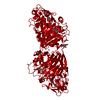
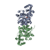
- Structure visualization
| Structure viewer | Molecule: MolmilJmol/JSmol |
|---|
- Downloads & links
-Download
| PDBx/mmCIF format | 3tze.cif.gz | 279.6 KB | Display | PDBx/mmCIF format |
|---|---|---|---|---|
| PDB format | pdb3tze.ent.gz | 225.7 KB | Display | PDB format |
| PDBx/mmJSON format | 3tze.json.gz | Tree view | PDBx/mmJSON format | |
| Others |  Other downloads Other downloads |
-Validation report
| Arichive directory | https://data.pdbj.org/pub/pdb/validation_reports/tz/3tzeftp://data.pdbj.org/pub/pdb/validation_reports/tz/3tze | HTTPS FTP |
|---|
-Related structure data
| Related structure data | 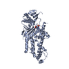 3sp1C  4e51C 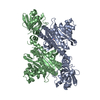 4ex5C 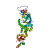 4g6zC  4griC  1ulhS S: Starting model for refinement C: citing same article ( |
|---|---|
| Similar structure data | |
| Other databases |


-Links
 PDBj
PDBj

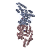
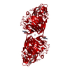
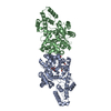
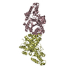
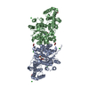
- Assembly
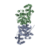
| Deposited unit | 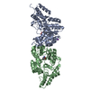
| ||||||||
|---|---|---|---|---|---|---|---|---|---|
| 1 |
| ||||||||
| Unit cell |
|
-Components
| #1: Protein | Mass: 46571.125 Da / Num. of mol.: 2 / Mutation: A149T Source method: isolated from a genetically manipulated source Source: (gene. exp.) Encephalitozoon cuniculi (fungus) / Strain: GB-M1 / Gene: ECU11_0530 / Production host:  #2: Chemical |   Type: L-peptide linking / Mass: 204.225 Da / Num. of mol.: 2 / Source method: obtained synthetically / Formula: C11H12N2O2 Type: L-peptide linking / Mass: 204.225 Da / Num. of mol.: 2 / Source method: obtained synthetically / Formula: C11H12N2O2#3: Chemical |   Mass: 39.098 Da / Num. of mol.: 2 / Source method: obtained synthetically / Formula: K Mass: 39.098 Da / Num. of mol.: 2 / Source method: obtained synthetically / Formula: K#4: Water | ChemComp-HOH / |  Mass: 18.015 Da / Num. of mol.: 61 / Source method: isolated from a natural source / Formula: H2O Mass: 18.015 Da / Num. of mol.: 61 / Source method: isolated from a natural source / Formula: H2O |
|---|
-Experimental details
-Experiment
| Experiment | Method: X-RAY DIFFRACTION / Number of used crystals: 1 |
|---|
- Sample preparation
| Crystal | Density Matthews: 2.04 Å3/Da / Density % sol: 39.56 % |
|---|---|
| Crystal grow | Temperature: 289 K / Method: vapor diffusion, sitting drop / pH: 7 Details: EncuA.00600.a.A1 PS00344 at 23.5 mg/mL against Wizard III screen from Emerald BioSystems, 20% PEG 3350, 0.2 M potassium nitrate, crystal tracking ID 221699a8, pH 7, VAPOR DIFFUSION, SITTING ...Details: EncuA.00600.a.A1 PS00344 at 23.5 mg/mL against Wizard III screen from Emerald BioSystems, 20% PEG 3350, 0.2 M potassium nitrate, crystal tracking ID 221699a8, pH 7, VAPOR DIFFUSION, SITTING DROP, temperature 289K |
-Data collection
| Diffraction | Mean temperature: 100 K | ||||||||||||||||||||||||||||||||||||||||||||||||||||||||||||||||||||||||||||||||||||||||||||||||||||||||||||||||||||||||||||||||||||||||||||||||||||||||||||||||||||||||
|---|---|---|---|---|---|---|---|---|---|---|---|---|---|---|---|---|---|---|---|---|---|---|---|---|---|---|---|---|---|---|---|---|---|---|---|---|---|---|---|---|---|---|---|---|---|---|---|---|---|---|---|---|---|---|---|---|---|---|---|---|---|---|---|---|---|---|---|---|---|---|---|---|---|---|---|---|---|---|---|---|---|---|---|---|---|---|---|---|---|---|---|---|---|---|---|---|---|---|---|---|---|---|---|---|---|---|---|---|---|---|---|---|---|---|---|---|---|---|---|---|---|---|---|---|---|---|---|---|---|---|---|---|---|---|---|---|---|---|---|---|---|---|---|---|---|---|---|---|---|---|---|---|---|---|---|---|---|---|---|---|---|---|---|---|---|---|---|---|---|
| Diffraction source | Source: SYNCHROTRON / Site: CLSI  / Beamline: 08ID-1 / Wavelength: 0.97949 Å / Beamline: 08ID-1 / Wavelength: 0.97949 Å | ||||||||||||||||||||||||||||||||||||||||||||||||||||||||||||||||||||||||||||||||||||||||||||||||||||||||||||||||||||||||||||||||||||||||||||||||||||||||||||||||||||||||
| Detector | Detector: CCD / Date: Sep 20, 2011 | ||||||||||||||||||||||||||||||||||||||||||||||||||||||||||||||||||||||||||||||||||||||||||||||||||||||||||||||||||||||||||||||||||||||||||||||||||||||||||||||||||||||||
| Radiation | Protocol: SINGLE WAVELENGTH / Monochromatic (M) / Laue (L): M / Scattering type: x-ray | ||||||||||||||||||||||||||||||||||||||||||||||||||||||||||||||||||||||||||||||||||||||||||||||||||||||||||||||||||||||||||||||||||||||||||||||||||||||||||||||||||||||||
| Radiation wavelength | Wavelength: 0.97949 Å / Relative weight: 1 | ||||||||||||||||||||||||||||||||||||||||||||||||||||||||||||||||||||||||||||||||||||||||||||||||||||||||||||||||||||||||||||||||||||||||||||||||||||||||||||||||||||||||
| Reflection | Resolution: 2.6→50 Å / Num. all: 24185 / Num. obs: 23335 / % possible obs: 96.5 % / Observed criterion σ(I): -3 / Redundancy: 4.8 % / Biso Wilson estimate: 48.833 Å2 / Rmerge(I) obs: 0.078 / Net I/σ(I): 14.71 | ||||||||||||||||||||||||||||||||||||||||||||||||||||||||||||||||||||||||||||||||||||||||||||||||||||||||||||||||||||||||||||||||||||||||||||||||||||||||||||||||||||||||
| Reflection shell | Diffraction-ID: 1
|
-Phasing
| Phasing | Method: molecular replacement | |||||||||
|---|---|---|---|---|---|---|---|---|---|---|
| Phasing MR | Model details: Phaser MODE: MR_AUTO
|
- Processing
| Software |
| |||||||||||||||||||||||||||||||||||||||||||||||||||||||||||||||||||||||||||||||||||||||||||||||||||||||||||||||||||||||||||||||||||||||||||||||||||||||||||||||||||||||||||||||||||||||||||||||||||||||||||||||||||||||||||||||||
|---|---|---|---|---|---|---|---|---|---|---|---|---|---|---|---|---|---|---|---|---|---|---|---|---|---|---|---|---|---|---|---|---|---|---|---|---|---|---|---|---|---|---|---|---|---|---|---|---|---|---|---|---|---|---|---|---|---|---|---|---|---|---|---|---|---|---|---|---|---|---|---|---|---|---|---|---|---|---|---|---|---|---|---|---|---|---|---|---|---|---|---|---|---|---|---|---|---|---|---|---|---|---|---|---|---|---|---|---|---|---|---|---|---|---|---|---|---|---|---|---|---|---|---|---|---|---|---|---|---|---|---|---|---|---|---|---|---|---|---|---|---|---|---|---|---|---|---|---|---|---|---|---|---|---|---|---|---|---|---|---|---|---|---|---|---|---|---|---|---|---|---|---|---|---|---|---|---|---|---|---|---|---|---|---|---|---|---|---|---|---|---|---|---|---|---|---|---|---|---|---|---|---|---|---|---|---|---|---|---|---|---|---|---|---|---|---|---|---|---|---|---|---|---|---|---|---|
| Refinement | Method to determine structure: MOLECULAR REPLACEMENT Starting model: PDB entry 1ULH Resolution: 2.6→50 Å / Cor.coef. Fo:Fc: 0.937 / Cor.coef. Fo:Fc free: 0.911 / WRfactor Rfree: 0.2232 / WRfactor Rwork: 0.1837 / Occupancy max: 1 / Occupancy min: 1 / FOM work R set: 0.8337 / SU B: 22.715 / SU ML: 0.234 / SU R Cruickshank DPI: 2.5214 / SU Rfree: 0.3291 / Cross valid method: THROUGHOUT / σ(F): 0 / ESU R Free: 0.329 / Stereochemistry target values: MAXIMUM LIKELIHOOD Details: U VALUES: WITH TLS ADDED HYDROGENS HAVE BEEN USED IF PRESENT IN THE INPUT
| |||||||||||||||||||||||||||||||||||||||||||||||||||||||||||||||||||||||||||||||||||||||||||||||||||||||||||||||||||||||||||||||||||||||||||||||||||||||||||||||||||||||||||||||||||||||||||||||||||||||||||||||||||||||||||||||||
| Solvent computation | Ion probe radii: 0.8 Å / Shrinkage radii: 0.8 Å / VDW probe radii: 1.2 Å / Solvent model: MASK | |||||||||||||||||||||||||||||||||||||||||||||||||||||||||||||||||||||||||||||||||||||||||||||||||||||||||||||||||||||||||||||||||||||||||||||||||||||||||||||||||||||||||||||||||||||||||||||||||||||||||||||||||||||||||||||||||
| Displacement parameters | Biso max: 88.86 Å2 / Biso mean: 43.2257 Å2 / Biso min: 23.67 Å2
| |||||||||||||||||||||||||||||||||||||||||||||||||||||||||||||||||||||||||||||||||||||||||||||||||||||||||||||||||||||||||||||||||||||||||||||||||||||||||||||||||||||||||||||||||||||||||||||||||||||||||||||||||||||||||||||||||
| Refinement step | Cycle: LAST / Resolution: 2.6→50 Å
| |||||||||||||||||||||||||||||||||||||||||||||||||||||||||||||||||||||||||||||||||||||||||||||||||||||||||||||||||||||||||||||||||||||||||||||||||||||||||||||||||||||||||||||||||||||||||||||||||||||||||||||||||||||||||||||||||
| Refine LS restraints |
| |||||||||||||||||||||||||||||||||||||||||||||||||||||||||||||||||||||||||||||||||||||||||||||||||||||||||||||||||||||||||||||||||||||||||||||||||||||||||||||||||||||||||||||||||||||||||||||||||||||||||||||||||||||||||||||||||
| LS refinement shell | Resolution: 2.6→2.667 Å / Total num. of bins used: 20
| |||||||||||||||||||||||||||||||||||||||||||||||||||||||||||||||||||||||||||||||||||||||||||||||||||||||||||||||||||||||||||||||||||||||||||||||||||||||||||||||||||||||||||||||||||||||||||||||||||||||||||||||||||||||||||||||||
| Refinement TLS params. | Method: refined / Refine-ID: X-RAY DIFFRACTION
| |||||||||||||||||||||||||||||||||||||||||||||||||||||||||||||||||||||||||||||||||||||||||||||||||||||||||||||||||||||||||||||||||||||||||||||||||||||||||||||||||||||||||||||||||||||||||||||||||||||||||||||||||||||||||||||||||
| Refinement TLS group |
|
