Movie
Movie Controller

[English] 日本語
 Yorodumi
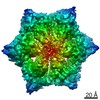
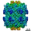
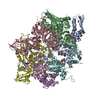
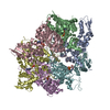
- PDB-6pp8: ClpX in ClpX-ClpP complex bound to substrate and ATP-gamma-S, class 1 -
+ Open data

- Basic information
| Entry | Database: PDB / ID: 6pp8 | |||||||||
|---|---|---|---|---|---|---|---|---|---|---|
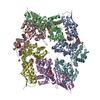
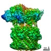
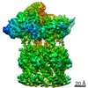
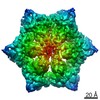
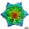
| Title | ClpX in ClpX-ClpP complex bound to substrate and ATP-gamma-S, class 1 | |||||||||
 Components Components |
| |||||||||
 Keywords Keywords | CHAPERONE / Protein degradation / AAA+ protease complex | |||||||||
| Function / homology |  Function and homology information Function and homology informationHslUV protease complex / endopeptidase Clp complex / ATP-dependent peptidase activity / protein unfolding / : / ATP-dependent protein folding chaperone / disordered domain specific binding / : / peptidase activity / protein folding ...HslUV protease complex / endopeptidase Clp complex / ATP-dependent peptidase activity / protein unfolding / : / ATP-dependent protein folding chaperone / disordered domain specific binding / : / peptidase activity / protein folding / protease binding / protein dimerization activity / cell division / ATP hydrolysis activity / zinc ion binding / ATP binding / identical protein binding / cytosol Similarity search - Function | |||||||||
| Biological species |  | |||||||||
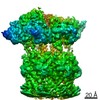
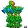
| Method | ELECTRON MICROSCOPY / single particle reconstruction / cryo EM / Resolution: 4.12 Å | |||||||||
 Authors Authors | Fei, X. / Jenni, S. / Harrison, S.C. / Sauer, R.T. | |||||||||
| Funding support |  United States, 2items United States, 2items
| |||||||||
 Citation Citation | Journal: Elife / Year: 2020 Title: Structures of the ATP-fueled ClpXP proteolytic machine bound to protein substrate. Authors: Xue Fei / Tristan A Bell / Simon Jenni / Benjamin M Stinson / Tania A Baker / Stephen C Harrison / Robert T Sauer / Abstract: ClpXP is an ATP-dependent protease in which the ClpX AAA+ motor binds, unfolds, and translocates specific protein substrates into the degradation chamber of ClpP. We present cryo-EM studies of the ...ClpXP is an ATP-dependent protease in which the ClpX AAA+ motor binds, unfolds, and translocates specific protein substrates into the degradation chamber of ClpP. We present cryo-EM studies of the enzyme that show how asymmetric hexameric rings of ClpX bind symmetric heptameric rings of ClpP and interact with protein substrates. Subunits in the ClpX hexamer assume a spiral conformation and interact with two-residue segments of substrate in the axial channel, as observed for other AAA+ proteases and protein-remodeling machines. Strictly sequential models of ATP hydrolysis and a power stroke that moves two residues of the substrate per translocation step have been inferred from these structural features for other AAA+ unfoldases, but biochemical and single-molecule biophysical studies indicate that ClpXP operates by a probabilistic mechanism in which five to eight residues are translocated for each ATP hydrolyzed. We propose structure-based models that could account for the functional results. | |||||||||
| History |
|
- Structure visualization
| Movie |
Movie viewer |
|---|---|
| Structure viewer | Molecule: MolmilJmol/JSmol |
- Downloads & links
-Download
| PDBx/mmCIF format | 6pp8.cif.gz | 665.9 KB | Display | PDBx/mmCIF format |
|---|---|---|---|---|
| PDB format | pdb6pp8.ent.gz | 560.4 KB | Display | PDB format |
| PDBx/mmJSON format | 6pp8.json.gz | Tree view | PDBx/mmJSON format | |
| Others |  Other downloads Other downloads |
-Validation report
| Arichive directory | https://data.pdbj.org/pub/pdb/validation_reports/pp/6pp8ftp://data.pdbj.org/pub/pdb/validation_reports/pp/6pp8 | HTTPS FTP |
|---|
-Related structure data
| Related structure data | 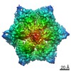 20422MC 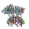 6po1C 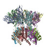 6po3C 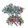 6podC 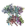 6posC 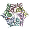 6pp5C 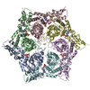 6pp6C 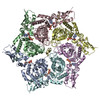 6pp7C 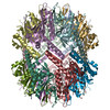 6ppeC C: citing same article ( M: map data used to model this data |
|---|---|
| Similar structure data |


-Links
 PDBj
PDBj


- Assembly
| Deposited unit | 
|
|---|---|
| 1 |
|
-Components
| #1: Protein | Mass: 39835.129 Da / Num. of mol.: 6 / Mutation: E185Q Source method: isolated from a genetically manipulated source Source: (gene. exp.) #2: Protein/peptide | | Mass: 770.943 Da / Num. of mol.: 1 Source method: isolated from a genetically manipulated source Source: (gene. exp.) #3: Chemical | ChemComp-AGS /   Mass: 523.247 Da / Num. of mol.: 5 / Source method: obtained synthetically / Formula: C10H16N5O12P3S / Comment: ATP-gamma-S, energy-carrying molecule analogue*YM Mass: 523.247 Da / Num. of mol.: 5 / Source method: obtained synthetically / Formula: C10H16N5O12P3S / Comment: ATP-gamma-S, energy-carrying molecule analogue*YM#4: Chemical | ChemComp-ADP / |   Mass: 427.201 Da / Num. of mol.: 1 / Source method: obtained synthetically / Formula: C10H15N5O10P2 / Comment: ADP, energy-carrying molecule*YM Mass: 427.201 Da / Num. of mol.: 1 / Source method: obtained synthetically / Formula: C10H15N5O10P2 / Comment: ADP, energy-carrying molecule*YMHas ligand of interest | N | Has protein modification | N | |
|---|
-Experimental details
-Experiment
| Experiment | Method: ELECTRON MICROSCOPY |
|---|---|
| EM experiment | Aggregation state: PARTICLE / 3D reconstruction method: single particle reconstruction |
- Sample preparation
| Component | Name: ClpX-ClpP-substrate-ATPrS / Type: COMPLEX / Entity ID: #1-#2 / Source: RECOMBINANT |
|---|---|
| Molecular weight | Experimental value: NO |
| Source (natural) | Organism: |
| Source (recombinant) | Organism: |
| Buffer solution | pH: 7.5 |
| Specimen | Embedding applied: NO / Shadowing applied: NO / Staining applied: NO / Vitrification applied: YES |
| Specimen support | Grid material: COPPER / Grid mesh size: 400 divisions/in. / Grid type: Quantifoil R1.2/1.3 |
| Vitrification | Instrument: GATAN CRYOPLUNGE 3 / Cryogen name: ETHANE / Humidity: 95 % / Chamber temperature: 298 K |
- Electron microscopy imaging
| Experimental equipment |  Model: Talos Arctica / Image courtesy: FEI Company |
|---|---|
| Microscopy | Model: FEI TECNAI ARCTICA |
| Electron gun | Electron source:  FIELD EMISSION GUN / Accelerating voltage: 200 kV / Illumination mode: OTHER FIELD EMISSION GUN / Accelerating voltage: 200 kV / Illumination mode: OTHER |
| Electron lens | Mode: BRIGHT FIELD / Nominal magnification: 36000 X / Nominal defocus min: -800 nm / Calibrated defocus max: -2500 nm / Alignment procedure: COMA FREE |
| Specimen holder | Cryogen: NITROGEN / Specimen holder model: FEI TITAN KRIOS AUTOGRID HOLDER |
| Image recording | Average exposure time: 60 sec. / Electron dose: 56 e/Å2 / Detector mode: SUPER-RESOLUTION / Film or detector model: GATAN K2 SUMMIT (4k x 4k) / Num. of grids imaged: 2 |
- Processing
| Software | Name: PHENIX / Version: 1.14_3260: / Classification: refinement | ||||||||||||||||||||||||||||||||||||
|---|---|---|---|---|---|---|---|---|---|---|---|---|---|---|---|---|---|---|---|---|---|---|---|---|---|---|---|---|---|---|---|---|---|---|---|---|---|
| EM software |
| ||||||||||||||||||||||||||||||||||||
| Image processing | Details: 2X binned and motioncor2 corrected | ||||||||||||||||||||||||||||||||||||
| CTF correction | Type: PHASE FLIPPING AND AMPLITUDE CORRECTION | ||||||||||||||||||||||||||||||||||||
| 3D reconstruction | Resolution: 4.12 Å / Resolution method: FSC 0.143 CUT-OFF / Num. of particles: 151652 / Symmetry type: POINT | ||||||||||||||||||||||||||||||||||||
| Atomic model building | PDB-ID: 3HWS Pdb chain-ID: A / Accession code: 3HWS / Source name: PDB / Type: experimental model | ||||||||||||||||||||||||||||||||||||
| Refine LS restraints |
|