Resolution: 2.27→96.99 Å / SU ML: 0.144 / Cross valid method: THROUGHOUT / ESU R: 0.267 / ESU R Free: 0.19 Details: Solved, built, and refined with Phenix (Phaser, Autobuild, Refine). "Polished" with PDB_REDO (Refmac).
Rfactor
Num. reflection
% reflection
Selection details
Rfree
0.20779
2402
4.8 %
RANDOM
Rwork
0.18111
-
-
-
obs
0.18243
47164
96.82 %
-
Solvent computation
Ion probe radii: 0.7 Å / Shrinkage radii: 0.7 Å / VDW probe radii: 1.1 Å
Displacement parameters
Biso mean: 32.552 Å2
Baniso -1
Baniso -2
Baniso -3
1-
0.2 Å2
0.1 Å2
0 Å2
2-
-
0.2 Å2
-0 Å2
3-
-
-
-0.66 Å2
Refinement step
Cycle: LAST / Resolution: 2.27→96.99 Å
Protein
Nucleic acid
Ligand
Solvent
Total
Num. atoms
7011
0
48
179
7238
LS refinement shell
Resolution: 2.272→2.331 Å
Rfactor
Num. reflection
% reflection
Rfree
0.296
189
5.3 %
Rwork
0.231
3559
-
obs
-
-
99.92 %
+
About Yorodumi
-
News
-
Feb 9, 2022. New format data for meta-information of EMDB entries
New format data for meta-information of EMDB entries
Version 3 of the EMDB header file is now the official format.
The previous official version 1.9 will be removed from the archive.
In the structure databanks used in Yorodumi, some data are registered as the other names, "COVID-19 virus" and "2019-nCoV". Here are the details of the virus and the list of structure data.
Jan 31, 2019. EMDB accession codes are about to change! (news from PDBe EMDB page)
EMDB accession codes are about to change! (news from PDBe EMDB page)
The allocation of 4 digits for EMDB accession codes will soon come to an end. Whilst these codes will remain in use, new EMDB accession codes will include an additional digit and will expand incrementally as the available range of codes is exhausted. The current 4-digit format prefixed with “EMD-” (i.e. EMD-XXXX) will advance to a 5-digit format (i.e. EMD-XXXXX), and so on. It is currently estimated that the 4-digit codes will be depleted around Spring 2019, at which point the 5-digit format will come into force.
The EM Navigator/Yorodumi systems omit the EMD- prefix.
Related info.:Q: What is EMD? / ID/Accession-code notation in Yorodumi/EM Navigator
Yorodumi is a browser for structure data from EMDB, PDB, SASBDB, etc.
This page is also the successor to EM Navigator detail page, and also detail information page/front-end page for Omokage search.
The word "yorodu" (or yorozu) is an old Japanese word meaning "ten thousand". "mi" (miru) is to see.
Related info.:EMDB / PDB / SASBDB / Comparison of 3 databanks / Yorodumi Search / Aug 31, 2016. New EM Navigator & Yorodumi / Yorodumi Papers / Jmol/JSmol / Function and homology information / Changes in new EM Navigator and Yorodumi
 Movie
Movie Controller

 Yorodumi
 Open data
 Basic information
 Components
 Keywords
 Function and homology information
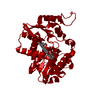
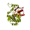
 Staphylococcus aureus (bacteria)
 X-RAY DIFFRACTION /
  Authors
 United States, 1items
  Citation
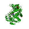
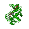
 Structure visualization
 Downloads & links
 Other downloads


 PDBj
PDBj
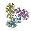
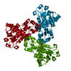
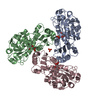
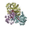
 Assembly
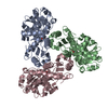
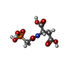
 Mass: 255.119 Da / Num. of mol.: 3 / Source method: obtained synthetically / Formula: C6H10NO8P / Feature type: SUBJECT OF INVESTIGATION
 Mass: 18.015 Da / Num. of mol.: 179 / Source method: isolated from a natural source / Formula: H2O
 Sample preparation
 Processing