 Movie
Movie Controller

[English] 日本語
 Yorodumi
- PDB-3l05: Crystal structure of N-acetyl-L-ornithine transcarbamylase E92S m... -
+ Open data

- Basic information
| Entry | Database: PDB / ID: 3l05 | |||||||||
|---|---|---|---|---|---|---|---|---|---|---|
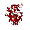
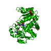
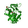
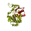
| Title | Crystal structure of N-acetyl-L-ornithine transcarbamylase E92S mutant complexed with carbamyl phosphate and N-succinyl-L-norvaline | |||||||||
 Components Components | N-acetylornithine carbamoyltransferase | |||||||||
 Keywords Keywords | TRANSFERASE / Transcarbamylase / Amino-acid biosynthesis / Arginine biosynthesis | |||||||||
| Function / homology |  Function and homology information Function and homology informationN-acetylornithine carbamoyltransferase / N-acetylornithine carbamoyltransferase activity / ornithine carbamoyltransferase activity / L-citrulline biosynthetic process / : / amino acid binding / cytoplasm Similarity search - Function | |||||||||
| Biological species |  Xanthomonas campestris pv. campestris (bacteria) Xanthomonas campestris pv. campestris (bacteria) | |||||||||
| Method |  X-RAY DIFFRACTION / MOLECULAR REPLACEMENT / Resolution: 2.8 Å X-RAY DIFFRACTION / MOLECULAR REPLACEMENT / Resolution: 2.8 Å | |||||||||
 Authors Authors | Shi, D. / Yu, X. / Allewell, N.M. / Tuchman, M. | |||||||||
 Citation Citation | Journal: Protein Sci. / Year: 2007 Title: A single mutation in the active site swaps the substrate specificity of N-acetyl-L-ornithine transcarbamylase and N-succinyl-L-ornithine transcarbamylase. Authors: Shi, D. / Yu, X. / Cabrera-Luque, J. / Chen, T.Y. / Roth, L. / Morizono, H. / Allewell, N.M. / Tuchman, M. | |||||||||
| History |
|
- Structure visualization
| Structure viewer | Molecule: MolmilJmol/JSmol |
|---|
- Downloads & links
-Download
| PDBx/mmCIF format | 3l05.cif.gz | 83.2 KB | Display | PDBx/mmCIF format |
|---|---|---|---|---|
| PDB format | pdb3l05.ent.gz | 61 KB | Display | PDB format |
| PDBx/mmJSON format | 3l05.json.gz | Tree view | PDBx/mmJSON format | |
| Others |  Other downloads Other downloads |
-Validation report
| Arichive directory | https://data.pdbj.org/pub/pdb/validation_reports/l0/3l05ftp://data.pdbj.org/pub/pdb/validation_reports/l0/3l05 | HTTPS FTP |
|---|
-Related structure data
| Related structure data |  2g7mC  3l02C  3l04C  3l06C  3kzoS S: Starting model for refinement C: citing same article ( |
|---|---|
| Similar structure data |


-Links
 PDBj
PDBj
- Assembly
| Deposited unit | 
| |||||||||
|---|---|---|---|---|---|---|---|---|---|---|
| 1 | 
| |||||||||
| Unit cell |
| |||||||||
| Components on special symmetry positions |
|
-Components
| #1: Protein | Mass: 40091.531 Da / Num. of mol.: 1 / Mutation: E92S Source method: isolated from a genetically manipulated source Source: (gene. exp.) Xanthomonas campestris pv. campestris (bacteria)Strain: ATCC 33913 / Gene: argF, argF', XCC2249 / Plasmid: pET28a / Production host: References: UniProt: Q8P8J2, N-acetylornithine carbamoyltransferase |
|---|---|
| #2: Chemical | ChemComp-SN0 /   Mass: 217.219 Da / Num. of mol.: 1 / Source method: obtained synthetically / Formula: C9H15NO5 Mass: 217.219 Da / Num. of mol.: 1 / Source method: obtained synthetically / Formula: C9H15NO5 |
| #3: Chemical | ChemComp-CP /   Mass: 141.020 Da / Num. of mol.: 1 / Source method: obtained synthetically / Formula: CH4NO5P Mass: 141.020 Da / Num. of mol.: 1 / Source method: obtained synthetically / Formula: CH4NO5P |
| #4: Chemical | ChemComp-SO4 /   Mass: 96.063 Da / Num. of mol.: 1 / Source method: obtained synthetically / Formula: SO4 Mass: 96.063 Da / Num. of mol.: 1 / Source method: obtained synthetically / Formula: SO4 |
| #5: Water | ChemComp-HOH /  Mass: 18.015 Da / Num. of mol.: 60 / Source method: isolated from a natural source / Formula: H2O Mass: 18.015 Da / Num. of mol.: 60 / Source method: isolated from a natural source / Formula: H2O |
-Experimental details
-Experiment
| Experiment | Method: X-RAY DIFFRACTION / Number of used crystals: 1 |
|---|
- Sample preparation
| Crystal | Density Matthews: 2.23 Å3/Da / Density % sol: 44.83 % |
|---|---|
| Crystal grow | Temperature: 291 K / Method: vapor diffusion, hanging drop / pH: 6 Details: Lithium sulfate, Bis-tris, PEG3350, pH 6.0, VAPOR DIFFUSION, HANGING DROP, temperature 291K |
-Data collection
| Diffraction | Mean temperature: 95 K |
|---|---|
| Diffraction source | Source: ROTATING ANODE / Type: RIGAKU RU300 / Wavelength: 1.54178 Å |
| Detector | Type: RIGAKU RAXIS IV / Detector: IMAGE PLATE / Date: Dec 27, 2005 |
| Radiation | Protocol: SINGLE WAVELENGTH / Monochromatic (M) / Laue (L): M / Scattering type: x-ray |
| Radiation wavelength | Wavelength: 1.54178 Å / Relative weight: 1 |
| Reflection | Resolution: 2.8→50 Å / Num. obs: 8953 / % possible obs: 99.8 % / Observed criterion σ(F): 0 / Observed criterion σ(I): 0 / Redundancy: 10.1 % / Biso Wilson estimate: 110.9 Å2 / Rmerge(I) obs: 0.153 / Χ2: 1.028 / Net I/σ(I): 15.8 |
| Reflection shell | Resolution: 2.8→2.9 Å / Redundancy: 9.7 % / Rmerge(I) obs: 0.787 / Mean I/σ(I) obs: 3.3 / Num. unique all: 875 / Χ2: 1.063 / % possible all: 100 |
- Processing
| Software |
| ||||||||||||||||||||||||||||||||||||
|---|---|---|---|---|---|---|---|---|---|---|---|---|---|---|---|---|---|---|---|---|---|---|---|---|---|---|---|---|---|---|---|---|---|---|---|---|---|
| Refinement | Method to determine structure: MOLECULAR REPLACEMENT Starting model: PDB entry 3KZO Resolution: 2.8→30.4 Å / Rfactor Rfree error: 0.012 / Occupancy max: 1 / Occupancy min: 0.33 / Data cutoff high absF: 2163796 / Data cutoff low absF: 0 / Isotropic thermal model: RESTRAINED / Cross valid method: THROUGHOUT / σ(F): 0 / Stereochemistry target values: Engh & Huber
| ||||||||||||||||||||||||||||||||||||
| Solvent computation | Solvent model: FLAT MODEL / Bsol: 29.582 Å2 / ksol: 0.328 e/Å3 | ||||||||||||||||||||||||||||||||||||
| Displacement parameters | Biso max: 70.96 Å2 / Biso mean: 38.489 Å2 / Biso min: 11.52 Å2
| ||||||||||||||||||||||||||||||||||||
| Refine analyze |
| ||||||||||||||||||||||||||||||||||||
| Refinement step | Cycle: LAST / Resolution: 2.8→30.4 Å
| ||||||||||||||||||||||||||||||||||||
| Refine LS restraints |
| ||||||||||||||||||||||||||||||||||||
| LS refinement shell | Resolution: 2.8→2.98 Å / Rfactor Rfree error: 0.04 / Total num. of bins used: 6
| ||||||||||||||||||||||||||||||||||||
| Xplor file |
|
