 Movie
Movie Controller

[English] 日本語
 Yorodumi
- PDB-6pl5: Structural coordination of polymerization and crosslinking by a p... -
+ Open data

- Basic information
| Entry | Database: PDB / ID: 6pl5 | ||||||
|---|---|---|---|---|---|---|---|
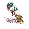
| Title | Structural coordination of polymerization and crosslinking by a peptidoglycan synthase complex | ||||||
 Components Components |
| ||||||
 Keywords Keywords | MEMBRANE PROTEIN / Peptidoglycan Glycosyltransferase / transmembrane protein / Shape Elongation Division and Sporulation / elongasome / Peptidoglycan transpeptidase | ||||||
| Function / homology |  Function and homology information Function and homology informationlipid-linked peptidoglycan transporter activity / peptidoglycan glycosyltransferase / peptidoglycan L,D-transpeptidase activity / peptidoglycan glycosyltransferase activity / cell division site / penicillin binding / peptidoglycan biosynthetic process / cell wall organization / regulation of cell shape / cell division / plasma membrane Similarity search - Function | ||||||
| Biological species |   Thermus thermophilus HB8 (bacteria) Thermus thermophilus HB8 (bacteria) | ||||||
| Method |  X-RAY DIFFRACTION / SYNCHROTRON / MOLECULAR REPLACEMENT / molecular replacement / Resolution: 3.5 Å X-RAY DIFFRACTION / SYNCHROTRON / MOLECULAR REPLACEMENT / molecular replacement / Resolution: 3.5 Å | ||||||
 Authors Authors | Sjodt, M. / Rohs, P.D.A. / Erlandson, S.C. / Zheng, S. / Rudner, D.Z. / Bernhardt, T.G. / Kruse, A.C. | ||||||
| Funding support |  United States, 1items United States, 1items
| ||||||
 Citation Citation | Journal: Nat Microbiol / Year: 2020 Title: Structural coordination of polymerization and crosslinking by a SEDS-bPBP peptidoglycan synthase complex. Authors: Sjodt, M. / Rohs, P.D.A. / Gilman, M.S.A. / Erlandson, S.C. / Zheng, S. / Green, A.G. / Brock, K.P. / Taguchi, A. / Kahne, D. / Walker, S. / Marks, D.S. / Rudner, D.Z. / Bernhardt, T.G. / Kruse, A.C. | ||||||
| History |
|
- Structure visualization
| Structure viewer | Molecule: MolmilJmol/JSmol |
|---|
- Downloads & links
-Download
| PDBx/mmCIF format | 6pl5.cif.gz | 371.9 KB | Display | PDBx/mmCIF format |
|---|---|---|---|---|
| PDB format | pdb6pl5.ent.gz | 303.5 KB | Display | PDB format |
| PDBx/mmJSON format | 6pl5.json.gz | Tree view | PDBx/mmJSON format | |
| Others |  Other downloads Other downloads |
-Validation report
| Arichive directory | https://data.pdbj.org/pub/pdb/validation_reports/pl/6pl5ftp://data.pdbj.org/pub/pdb/validation_reports/pl/6pl5 | HTTPS FTP |
|---|
-Related structure data
| Related structure data |  6pl6C  3lo7S  6barS S: Starting model for refinement C: citing same article ( |
|---|---|
| Similar structure data |


-Links
 PDBj
PDBj- Assembly
| Deposited unit | 
| ||||||||
|---|---|---|---|---|---|---|---|---|---|
| 1 |
| ||||||||
| Unit cell |
|
-Components
| #1: Protein | Mass: 40597.852 Da / Num. of mol.: 1 Source method: isolated from a genetically manipulated source Source: (gene. exp.) Thermus thermophilus HB8 (bacteria) / Strain: HB8 / ATCC 27634 / DSM 579 / Gene: rodA, TTHA1241 / Plasmid: pCOLA / Production host: References: UniProt: Q5SIX3, peptidoglycan glycosyltransferase |
|---|---|
| #2: Protein | Mass: 65452.074 Da / Num. of mol.: 1 Source method: isolated from a genetically manipulated source Source: (gene. exp.) Thermus thermophilus HB8 (bacteria) / Strain: HB8 / ATCC 27634 / DSM 579 / Gene: TTHA1191 / Production host: |
| #3: Protein/peptide | Mass: 954.168 Da / Num. of mol.: 1 Source method: isolated from a genetically manipulated source Source: (gene. exp.) Thermus thermophilus HB8 (bacteria) / Production host: |
| Has protein modification | Y |
-Experimental details
-Experiment
| Experiment | Method: X-RAY DIFFRACTION / Number of used crystals: 1 |
|---|
- Sample preparation
| Crystal | Density Matthews: 5.2 Å3/Da / Density % sol: 76.37 % |
|---|---|
| Crystal grow | Temperature: 293 K / Method: lipidic cubic phase / pH: 6.4 Details: Protein complex was reconstituted with monoolein. Crystals were grown in 100 mM MES pH 5.8-6.5, 100 mM Li2SO4, 40-50% PEG 300 PH range: 5.8-6.5 |
-Data collection
| Diffraction | Mean temperature: 100 K / Serial crystal experiment: N |
|---|---|
| Diffraction source | Source: SYNCHROTRON / Site: APS / Beamline: 23-ID-B / Wavelength: 1.033 Å |
| Detector | Type: DECTRIS EIGER X 16M / Detector: PIXEL / Date: Mar 22, 2018 |
| Radiation | Protocol: SINGLE WAVELENGTH / Monochromatic (M) / Laue (L): M / Scattering type: x-ray |
| Radiation wavelength | Wavelength: 1.033 Å / Relative weight: 1 |
| Reflection | Resolution: 3.5→44.766 Å / Num. obs: 50777 / % possible obs: 99.3 % / Redundancy: 6.11 % / Biso Wilson estimate: 155.59 Å2 / CC1/2: 0.999 / Rmerge(I) obs: 0.159 / Net I/σ(I): 8.15 |
| Reflection shell | Resolution: 3.5→3.59 Å / Redundancy: 6.11 % / Mean I/σ(I) obs: 0.62 / Num. unique obs: 2071 / CC1/2: 0.334 / % possible all: 99.8 |
-Phasing
| Phasing | Method: molecular replacement |
|---|
- Processing
| Software |
| ||||||||||||||||||||||||||||||||||||||||||||||||||||||||||||||||||||||||||||||||||||||||||||||||||||||||||||||||||||||||||||||||||||||||||||||||||||||||||||||||||||||||||||||||||||||||||||||||||||||||||||||||||||||||||||||||||||||||||||||||||||||||||
|---|---|---|---|---|---|---|---|---|---|---|---|---|---|---|---|---|---|---|---|---|---|---|---|---|---|---|---|---|---|---|---|---|---|---|---|---|---|---|---|---|---|---|---|---|---|---|---|---|---|---|---|---|---|---|---|---|---|---|---|---|---|---|---|---|---|---|---|---|---|---|---|---|---|---|---|---|---|---|---|---|---|---|---|---|---|---|---|---|---|---|---|---|---|---|---|---|---|---|---|---|---|---|---|---|---|---|---|---|---|---|---|---|---|---|---|---|---|---|---|---|---|---|---|---|---|---|---|---|---|---|---|---|---|---|---|---|---|---|---|---|---|---|---|---|---|---|---|---|---|---|---|---|---|---|---|---|---|---|---|---|---|---|---|---|---|---|---|---|---|---|---|---|---|---|---|---|---|---|---|---|---|---|---|---|---|---|---|---|---|---|---|---|---|---|---|---|---|---|---|---|---|---|---|---|---|---|---|---|---|---|---|---|---|---|---|---|---|---|---|---|---|---|---|---|---|---|---|---|---|---|---|---|---|---|---|---|---|---|---|---|---|---|---|---|---|---|---|---|---|---|---|
| Refinement | Method to determine structure: MOLECULAR REPLACEMENT Starting model: 6BAR,3LO7 Resolution: 3.5→44.766 Å / SU ML: 0.67 / Cross valid method: FREE R-VALUE / σ(F): 1.33 / Phase error: 38.31
| ||||||||||||||||||||||||||||||||||||||||||||||||||||||||||||||||||||||||||||||||||||||||||||||||||||||||||||||||||||||||||||||||||||||||||||||||||||||||||||||||||||||||||||||||||||||||||||||||||||||||||||||||||||||||||||||||||||||||||||||||||||||||||
| Solvent computation | Shrinkage radii: 0.9 Å / VDW probe radii: 1.11 Å | ||||||||||||||||||||||||||||||||||||||||||||||||||||||||||||||||||||||||||||||||||||||||||||||||||||||||||||||||||||||||||||||||||||||||||||||||||||||||||||||||||||||||||||||||||||||||||||||||||||||||||||||||||||||||||||||||||||||||||||||||||||||||||
| Displacement parameters | Biso max: 419.99 Å2 / Biso mean: 175.8364 Å2 / Biso min: 75.51 Å2 | ||||||||||||||||||||||||||||||||||||||||||||||||||||||||||||||||||||||||||||||||||||||||||||||||||||||||||||||||||||||||||||||||||||||||||||||||||||||||||||||||||||||||||||||||||||||||||||||||||||||||||||||||||||||||||||||||||||||||||||||||||||||||||
| Refinement step | Cycle: final / Resolution: 3.5→44.766 Å
| ||||||||||||||||||||||||||||||||||||||||||||||||||||||||||||||||||||||||||||||||||||||||||||||||||||||||||||||||||||||||||||||||||||||||||||||||||||||||||||||||||||||||||||||||||||||||||||||||||||||||||||||||||||||||||||||||||||||||||||||||||||||||||
| LS refinement shell | Refine-ID: X-RAY DIFFRACTION / Rfactor Rfree error: 0 / Total num. of bins used: 24
| ||||||||||||||||||||||||||||||||||||||||||||||||||||||||||||||||||||||||||||||||||||||||||||||||||||||||||||||||||||||||||||||||||||||||||||||||||||||||||||||||||||||||||||||||||||||||||||||||||||||||||||||||||||||||||||||||||||||||||||||||||||||||||
| Refinement TLS params. | Method: refined / Refine-ID: X-RAY DIFFRACTION
| ||||||||||||||||||||||||||||||||||||||||||||||||||||||||||||||||||||||||||||||||||||||||||||||||||||||||||||||||||||||||||||||||||||||||||||||||||||||||||||||||||||||||||||||||||||||||||||||||||||||||||||||||||||||||||||||||||||||||||||||||||||||||||
| Refinement TLS group |
|
