 Movie
Movie Controller

[English] 日本語
 Yorodumi
- PDB-6piz: Crystal structure of HCV NS3/4A D168A protease in complex with P4... -
+ Open data

- Basic information
| Entry | Database: PDB / ID: 6piz | ||||||||||||
|---|---|---|---|---|---|---|---|---|---|---|---|---|---|
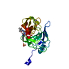
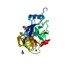
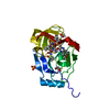
| Title | Crystal structure of HCV NS3/4A D168A protease in complex with P4-1 (NR02-24) | ||||||||||||
 Components Components | NS3/4A protease | ||||||||||||
 Keywords Keywords | hydrolase/hydrolase inhibitor / NS3/4a Protease / Hepatitis C virus / Drug Resistance / Protease inhibitor / HYDROLASE-HYDROLASE Inhibitor complex / HYDROLASE | ||||||||||||
| Function / homology |  Function and homology information Function and homology informationsymbiont-mediated transformation of host cell / host cell membrane / serine-type peptidase activity / viral capsid / host cell / RNA helicase activity / symbiont entry into host cell / virion attachment to host cell / virion membrane / proteolysis / metal ion binding Similarity search - Function | ||||||||||||
| Biological species |  Hepacivirus C Hepacivirus C | ||||||||||||
| Method |  X-RAY DIFFRACTION / MOLECULAR REPLACEMENT / Resolution: 1.89 Å X-RAY DIFFRACTION / MOLECULAR REPLACEMENT / Resolution: 1.89 Å | ||||||||||||
 Authors Authors | Zephyr, J. / Schiffer, C.A. | ||||||||||||
| Funding support |  United States, 3items United States, 3items
| ||||||||||||
 Citation Citation | Journal: Mbio / Year: 2020 Title: Avoiding Drug Resistance by Substrate Envelope-Guided Design: Toward Potent and Robust HCV NS3/4A Protease Inhibitors. Authors: Matthew, A.N. / Zephyr, J. / Nageswara Rao, D. / Henes, M. / Kamran, W. / Kosovrasti, K. / Hedger, A.K. / Lockbaum, G.J. / Timm, J. / Ali, A. / Kurt Yilmaz, N. / Schiffer, C.A. | ||||||||||||
| History |
|
- Structure visualization
| Structure viewer | Molecule: MolmilJmol/JSmol |
|---|
- Downloads & links
-Download
| PDBx/mmCIF format | 6piz.cif.gz | 92 KB | Display | PDBx/mmCIF format |
|---|---|---|---|---|
| PDB format | pdb6piz.ent.gz | 67.9 KB | Display | PDB format |
| PDBx/mmJSON format | 6piz.json.gz | Tree view | PDBx/mmJSON format | |
| Others |  Other downloads Other downloads |
-Validation report
| Arichive directory | https://data.pdbj.org/pub/pdb/validation_reports/pi/6pizftp://data.pdbj.org/pub/pdb/validation_reports/pi/6piz | HTTPS FTP |
|---|
-Related structure data
| Related structure data |  6piuC  6pivC  6piwC 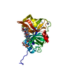 6pixC 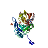 6piyC  6pj0C  6pj1C  6pj2C  6ue3C  5vojS S: Starting model for refinement C: citing same article ( |
|---|---|
| Similar structure data |


-Links
 PDBj
PDBj
- Assembly
| Deposited unit | 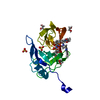
| ||||||||
|---|---|---|---|---|---|---|---|---|---|
| 1 |
| ||||||||
| Unit cell |
|
-Components
-Protein , 1 types, 1 molecules A
| #1: Protein | Mass: 23486.564 Da / Num. of mol.: 1 / Mutation: D168A Source method: isolated from a genetically manipulated source Source: (gene. exp.) Hepacivirus C / Production host:  |
|---|
-Non-polymers , 6 types, 187 molecules 


| #2: Chemical |  Mass: 96.063 Da / Num. of mol.: 2 / Source method: obtained synthetically / Formula: SO4 Mass: 96.063 Da / Num. of mol.: 2 / Source method: obtained synthetically / Formula: SO4#3: Chemical | ChemComp-ON4 / |  Mass: 752.877 Da / Num. of mol.: 1 / Source method: obtained synthetically / Formula: C37H48N6O9S Mass: 752.877 Da / Num. of mol.: 1 / Source method: obtained synthetically / Formula: C37H48N6O9S#4: Chemical | ChemComp-GOL / |  Mass: 92.094 Da / Num. of mol.: 1 / Source method: obtained synthetically / Formula: C3H8O3 Mass: 92.094 Da / Num. of mol.: 1 / Source method: obtained synthetically / Formula: C3H8O3#5: Chemical | ChemComp-EDO /  Mass: 62.068 Da / Num. of mol.: 4 / Source method: obtained synthetically / Formula: C2H6O2 Mass: 62.068 Da / Num. of mol.: 4 / Source method: obtained synthetically / Formula: C2H6O2#6: Chemical | ChemComp-ZN / |  Mass: 65.409 Da / Num. of mol.: 1 / Source method: isolated from a natural source / Formula: Zn Mass: 65.409 Da / Num. of mol.: 1 / Source method: isolated from a natural source / Formula: Zn#7: Water | ChemComp-HOH / | Mass: 18.015 Da / Num. of mol.: 178 / Source method: isolated from a natural source / Formula: H2O |
|---|
-Details
| Has ligand of interest | Y |
|---|
-Experimental details
-Experiment
| Experiment | Method: X-RAY DIFFRACTION / Number of used crystals: 1 |
|---|
- Sample preparation
| Crystal | Density Matthews: 2.04 Å3/Da / Density % sol: 39.63 % |
|---|---|
| Crystal grow | Temperature: 298 K / Method: vapor diffusion, hanging drop / pH: 6.5 Details: 100 mM MES Buffer pH 6.5, 4% (W/V) Ammonium Sulfate, 20-26% PEG 3350 |
-Data collection
| Diffraction | Mean temperature: 100 K / Serial crystal experiment: N |
|---|---|
| Diffraction source | Source: ROTATING ANODE / Type: RIGAKU MICROMAX-007 HF / Wavelength: 1.54178 Å |
| Detector | Type: RIGAKU SATURN 944 / Detector: CCD / Date: Dec 3, 2018 |
| Radiation | Protocol: SINGLE WAVELENGTH / Monochromatic (M) / Laue (L): M / Scattering type: x-ray |
| Radiation wavelength | Wavelength: 1.54178 Å / Relative weight: 1 |
| Reflection | Resolution: 1.89→50 Å / Num. obs: 15981 / % possible obs: 99.7 % / Redundancy: 6.8 % / Net I/σ(I): 20.04 |
| Reflection shell | Resolution: 1.89→1.92 Å |
- Processing
| Software |
| |||||||||||||||||||||||||||||||||||||||||||||||||
|---|---|---|---|---|---|---|---|---|---|---|---|---|---|---|---|---|---|---|---|---|---|---|---|---|---|---|---|---|---|---|---|---|---|---|---|---|---|---|---|---|---|---|---|---|---|---|---|---|---|---|
| Refinement | Method to determine structure: MOLECULAR REPLACEMENT Starting model: 5VOJ Resolution: 1.89→23.98 Å / SU ML: 0.13 / Cross valid method: FREE R-VALUE / σ(F): 1.34 / Phase error: 18.95
| |||||||||||||||||||||||||||||||||||||||||||||||||
| Solvent computation | Shrinkage radii: 0.9 Å / VDW probe radii: 1.11 Å | |||||||||||||||||||||||||||||||||||||||||||||||||
| Refinement step | Cycle: LAST / Resolution: 1.89→23.98 Å
| |||||||||||||||||||||||||||||||||||||||||||||||||
| Refine LS restraints |
| |||||||||||||||||||||||||||||||||||||||||||||||||
| LS refinement shell |
|
