 Movie
Movie Controller


+ Open data

- Basic information
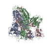
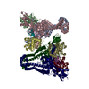
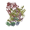
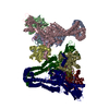
| Entry | Database: PDB / ID: 6pe5 | |||||||||
|---|---|---|---|---|---|---|---|---|---|---|
| Title | Yeast Vo motor in complex with 2 VopQ molecules | |||||||||
 Components Components |
| |||||||||
 Keywords Keywords | TRANSPORT PROTEIN / complex | |||||||||
| Function / homology |  Function and homology information Function and homology informationcell wall mannoprotein biosynthetic process / ATPase-coupled ion transmembrane transporter activity / cellular response to alkaline pH / protein localization to vacuolar membrane / Insulin receptor recycling / Transferrin endocytosis and recycling / polyphosphate metabolic process / ROS and RNS production in phagocytes / Amino acids regulate mTORC1 / Golgi lumen acidification ...cell wall mannoprotein biosynthetic process / ATPase-coupled ion transmembrane transporter activity / cellular response to alkaline pH / protein localization to vacuolar membrane / Insulin receptor recycling / Transferrin endocytosis and recycling / polyphosphate metabolic process / ROS and RNS production in phagocytes / Amino acids regulate mTORC1 / Golgi lumen acidification / P-type proton-exporting transporter activity / vacuolar transport / vacuolar proton-transporting V-type ATPase, V0 domain / endosomal lumen acidification / vacuole organization / protein targeting to vacuole / proton-transporting V-type ATPase complex / fungal-type vacuole / vacuolar proton-transporting V-type ATPase complex / cellular hyperosmotic response / vacuolar acidification / fungal-type vacuole membrane / phosphatidylinositol-3,5-bisphosphate binding / proton transmembrane transporter activity / proton-transporting ATPase activity, rotational mechanism / intracellular copper ion homeostasis / Neutrophil degranulation / RNA endonuclease activity / proton transmembrane transport / cell periphery / transmembrane transport / endocytosis / ATPase binding / protein-containing complex assembly / intracellular iron ion homeostasis / membrane raft / Golgi membrane / endoplasmic reticulum membrane / membrane Similarity search - Function | |||||||||
| Biological species |   Vibrio parahaemolyticus (bacteria) Vibrio parahaemolyticus (bacteria) | |||||||||
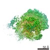
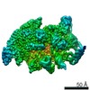
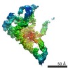
| Method | ELECTRON MICROSCOPY / single particle reconstruction / cryo EM / Resolution: 3.2 Å | |||||||||
 Authors Authors | Peng, W. / Li, Y. / Tomchick, D.R. / Orth, K. | |||||||||
| Funding support |  United States, 2items United States, 2items
| |||||||||
 Citation Citation | Journal: Nat Struct Mol Biol / Year: 2020 Title: A distinct inhibitory mechanism of the V-ATPase by Vibrio VopQ revealed by cryo-EM. Authors: Wei Peng / Amanda K Casey / Jessie Fernandez / Emily M Carpinone / Kelly A Servage / Zhe Chen / Yang Li / Diana R Tomchick / Vincent J Starai / Kim Orth / Abstract: The Vibrio parahaemolyticus T3SS effector VopQ targets host-cell V-ATPase, resulting in blockage of autophagic flux and neutralization of acidic compartments. Here, we report the cryo-EM structure of ...The Vibrio parahaemolyticus T3SS effector VopQ targets host-cell V-ATPase, resulting in blockage of autophagic flux and neutralization of acidic compartments. Here, we report the cryo-EM structure of VopQ bound to the V subcomplex of the V-ATPase. VopQ inserts into membranes and forms an unconventional pore while binding directly to subunit c of the V-ATPase membrane-embedded subcomplex V. We show that VopQ arrests yeast growth in vivo by targeting the immature V subcomplex in the endoplasmic reticulum (ER), thus providing insight into the observation that VopQ kills cells in the absence of a functional V-ATPase. VopQ is a bacterial effector that has been discovered to inhibit a host-membrane megadalton complex by coincidentally binding its target, inserting into a membrane and disrupting membrane potential. Collectively, our results reveal a mechanism by which bacterial effectors modulate host cell biology and provide an invaluable tool for future studies on V-ATPase-mediated membrane fusion and autophagy. | |||||||||
| History |
|
- Structure visualization
| Movie |
Movie viewer |
|---|---|
| Structure viewer | Molecule: MolmilJmol/JSmol |
- Downloads & links
-Download
| PDBx/mmCIF format | 6pe5.cif.gz | 616.4 KB | Display | PDBx/mmCIF format |
|---|---|---|---|---|
| PDB format | pdb6pe5.ent.gz | 500.1 KB | Display | PDB format |
| PDBx/mmJSON format | 6pe5.json.gz | Tree view | PDBx/mmJSON format | |
| Others |  Other downloads Other downloads |
-Validation report
| Summary document | 6pe5_validation.pdf.gz | 1.1 MB | Display | wwPDB validaton report |
|---|---|---|---|---|
| Full document | 6pe5_full_validation.pdf.gz | 1.1 MB | Display | |
| Data in XML | 6pe5_validation.xml.gz | 86.7 KB | Display | |
| Data in CIF | 6pe5_validation.cif.gz | 132.8 KB | Display | |
| Arichive directory | https://data.pdbj.org/pub/pdb/validation_reports/pe/6pe5ftp://data.pdbj.org/pub/pdb/validation_reports/pe/6pe5 | HTTPS FTP |
-Related structure data
| Related structure data | 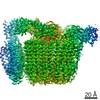 20323MC  6pe4C M: map data used to model this data C: citing same article ( |
|---|---|
| Similar structure data |

-Links
 PDBj
PDBj


- Assembly
| Deposited unit | 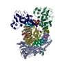
|
|---|---|
| 1 |
|
-Components
-V-type proton ATPase subunit ... , 6 types, 13 molecules ADEGHIJKLMNOP
| #1: Protein | Mass: 115126.023 Da / Num. of mol.: 1 / Source method: isolated from a natural source Source: (natural) Strain: ATCC 204508 / S288c / References: UniProt: P32563 |
|---|---|
| #3: Protein | Mass: 39822.484 Da / Num. of mol.: 1 / Source method: isolated from a natural source Source: (natural) Strain: ATCC 204508 / S288c / References: UniProt: P32366 |
| #4: Protein | Mass: 8387.065 Da / Num. of mol.: 1 / Source method: isolated from a natural source Source: (natural) Strain: ATCC 204508 / S288c / References: UniProt: Q3E7B6 |
| #6: Protein | Mass: 22610.641 Da / Num. of mol.: 1 / Source method: isolated from a natural source Source: (natural) Strain: ATCC 204508 / S288c / References: UniProt: P23968 |
| #7: Protein | Mass: 17046.361 Da / Num. of mol.: 1 / Source method: isolated from a natural source Source: (natural) Strain: ATCC 204508 / S288c / References: UniProt: P32842 |
| #8: Protein | Mass: 16357.501 Da / Num. of mol.: 8 / Source method: isolated from a natural source Source: (natural) Strain: ATCC 204508 / S288c / References: UniProt: P25515 |
-Protein , 3 types, 4 molecules BFQR
| #2: Protein | Mass: 29694.885 Da / Num. of mol.: 1 / Source method: isolated from a natural source Source: (natural) Strain: ATCC 204508 / S288c / References: UniProt: P53262 |
|---|---|
| #5: Protein | Mass: 9369.934 Da / Num. of mol.: 1 / Source method: isolated from a natural source Source: (natural) Strain: ATCC 204508 / S288c / References: UniProt: P0C5R9 |
| #9: Protein | Mass: 55424.668 Da / Num. of mol.: 2 Source method: isolated from a genetically manipulated source Source: (gene. exp.) Vibrio parahaemolyticus (bacteria) / Gene: ACS91_23375 / Production host: |
-Experimental details
-Experiment
| Experiment | Method: ELECTRON MICROSCOPY |
|---|---|
| EM experiment | Aggregation state: PARTICLE / 3D reconstruction method: single particle reconstruction |
- Sample preparation
| Component | Name: Yeast Vo motor in complex with 2 VopQ molecules / Type: COMPLEX / Entity ID: all / Source: RECOMBINANT |
|---|---|
| Molecular weight | Value: 0.46 MDa / Experimental value: NO |
| Source (natural) | Organism: |
| Source (recombinant) | Organism: |
| Buffer solution | pH: 7.4 |
| Specimen | Embedding applied: NO / Shadowing applied: NO / Staining applied: NO / Vitrification applied: YES |
| Vitrification | Cryogen name: ETHANE |
- Electron microscopy imaging
| Experimental equipment |  Model: Titan Krios / Image courtesy: FEI Company |
|---|---|
| Microscopy | Model: FEI TITAN KRIOS |
| Electron gun | Electron source:  FIELD EMISSION GUN / Accelerating voltage: 300 kV / Illumination mode: FLOOD BEAM FIELD EMISSION GUN / Accelerating voltage: 300 kV / Illumination mode: FLOOD BEAM |
| Electron lens | Mode: BRIGHT FIELD |
| Image recording | Electron dose: 50 e/Å2 / Detector mode: SUPER-RESOLUTION / Film or detector model: GATAN K2 SUMMIT (4k x 4k) |
- Processing
| CTF correction | Type: NONE |
|---|---|
| 3D reconstruction | Resolution: 3.2 Å / Resolution method: FSC 0.143 CUT-OFF / Num. of particles: 72837 / Symmetry type: POINT |
